ADEM on MRI: how Monitraq's AI read widespread white matter change in a child
Acute disseminated encephalomyelitis (ADEM) on MRI can look like a long list of other white matter diseases. In this child's brain scan the white matter was bright almost everywhere. Here is how Monitraq's AI worked through it.
MRI findings
A child had a brain MRI with T2, FLAIR, diffusion, susceptibility and T1 images before and after contrast. No clinical history came with the scan.
The cerebral white matter was diffusely bright on T2 and FLAIR, on both sides and largely symmetric. The change was confluent, ran through the frontal, parietal, occipital and temporal lobes, reached the subcortical white matter in places, and involved the front (genu) and back (splenium) of the corpus callosum. The deep grey nuclei, brainstem and cerebellar white matter were spared. There was no mass effect and the ventricles were normal in size.
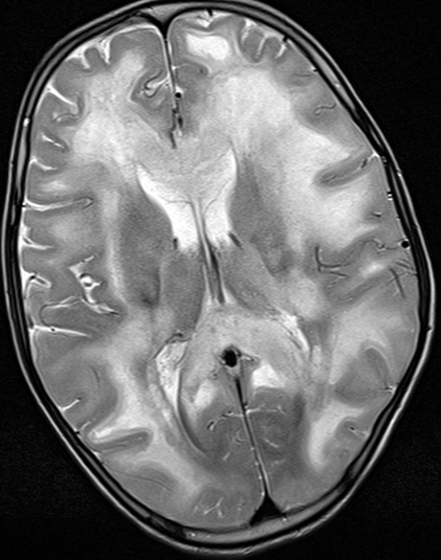
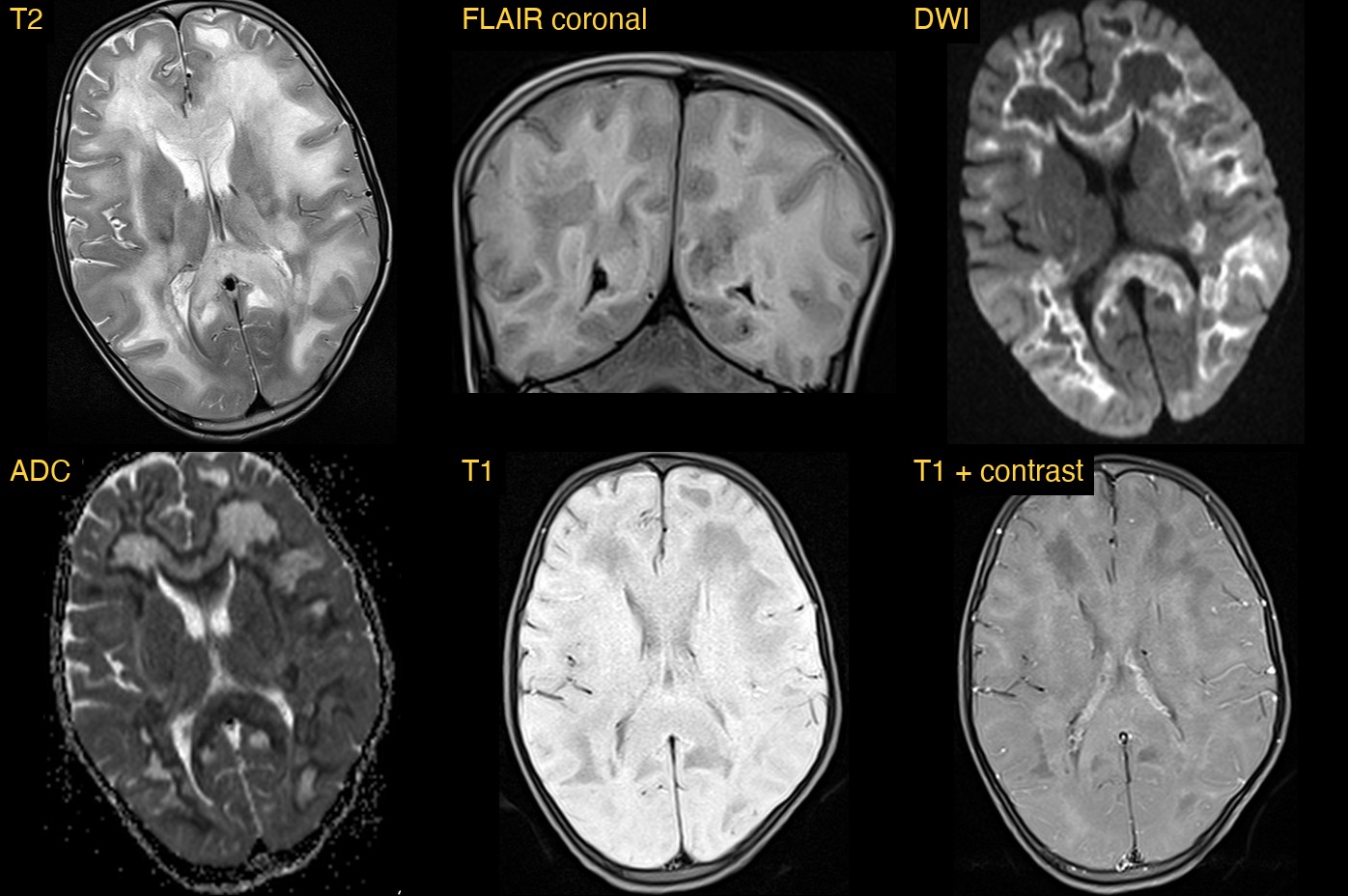
How Monitraq's AI read the scan
- It mapped the extent: diffuse, largely symmetric, confluent T2/FLAIR change in the periventricular, deep and partly subcortical white matter of all cerebral lobes, including the genu and splenium of the corpus callosum.
- It read diffusion in two parts: a peripheral band at the subcortical margin and in the corpus callosum showed restricted diffusion, a sign of active disease, while the central deep white matter had raised ADC with patchy partial FLAIR suppression.
- It noted what was spared: the deep grey nuclei, brainstem and cerebellar white matter, with no mass effect and normal-sized ventricles.
- It checked enhancement: the white matter was dark on T1 with no convincing enhancement after contrast.
- It gave a ranked differential: its favoured diagnosis was ADEM or MOG antibody-associated disease. The main inherited alternative was vanishing white matter disease, raised by the central rarefaction; X-linked adrenoleukodystrophy and metachromatic leukodystrophy were less likely.
- It flagged the case as extensive white matter disease with active restricted diffusion in a child, and listed next steps: the clinical story (tempo of onset, recent infection or vaccination, encephalopathy, regression), serum MOG and aquaporin-4 antibodies, CSF analysis, a metabolic screen, MR spectroscopy, spine MRI and a follow-up MRI.
Dr Priti, the radiologist who reviewed this case, reported acute disseminated encephalomyelitis (ADEM).
What is acute disseminated encephalomyelitis?
ADEM is an immune-mediated inflammatory disease of the brain and spinal cord that damages myelin, the insulating layer around nerve fibres. It is most common in children. It often follows an infection or, less often, a vaccination, and it usually happens as a single episode.
Children typically become unwell over days, with encephalopathy, meaning confusion, drowsiness or behaviour change, along with signs that point to several parts of the nervous system at once. Some children with ADEM have antibodies to myelin oligodendrocyte glycoprotein (MOG), which is why testing for MOG antibodies is part of the work-up.
Key MRI signs of ADEM
- Multiple large, poorly marginated T2/FLAIR bright lesions in the cerebral white matter, often on both sides but not always symmetric.
- Frequent involvement of the subcortical white matter and the junction with the cortex.
- The deep grey matter (thalami, basal ganglia), brainstem, cerebellum and spinal cord may also be involved.
- Lesions tend to be of a similar age, in keeping with a single episode.
- Enhancement is variable and may be absent.
- On follow-up, lesions usually improve or resolve.
Differential diagnosis
Widespread white matter change in a child has a long list of causes. Inflammatory causes include ADEM, MOG antibody-associated disease and, less often in young children, multiple sclerosis. Inherited white matter diseases, the leukodystrophies, can look similar on a single scan. Clues that favour a leukodystrophy include strict symmetry, a slow course over months, and tissue that is breaking down into fluid, as in vanishing white matter disease. That is why the AI weighed the central rarefaction carefully and asked for the clinical story, antibody tests, a metabolic screen and a follow-up scan.
Frequently asked questions
Which MRI sequence shows ADEM best?
T2 and FLAIR show the extent of the lesions best. DWI and ADC help show which areas are active, and post-contrast T1 shows any enhancement.
How is ADEM different from multiple sclerosis on MRI?
ADEM lesions are usually large, ill-defined and of similar age, often with deep grey matter involvement. Multiple sclerosis lesions are typically smaller, ovoid and periventricular, with new lesions appearing over time.
Why repeat the MRI in suspected ADEM?
ADEM lesions usually improve over the following months. A follow-up scan helps to confirm the diagnosis and to separate it from a progressive white matter disease.
A draft for the radiologist
Diffuse white matter change in a child is a pattern, not a diagnosis. A useful report says what is involved, what is spared and how diffusion behaves, then ranks the causes and lists the tests that will separate them. Monitraq's AI is built to do exactly that, and to show its reasoning so the radiologist can check it quickly.
It gives a draft, not a diagnosis. The AI flags, describes and suggests. A radiologist confirms, corrects and signs.
Monitraq's AI produces draft reports to support qualified radiologists. It is not a diagnosis and is not intended for use by patients. All case details have been removed or generalised to protect patient privacy, and the images have been cropped and stripped of identifying information.