A towering NAA peak: how Monitraq's AI recognised Canavan disease
A young child had a brain MRI with MR spectroscopy. The images showed widespread abnormal white matter, a picture shared by many rare conditions. The spectrum showed one peak far too tall. Here is how Monitraq's AI put the two together.
The scan
The study included T2, FLAIR, diffusion (DWI with an ADC map), susceptibility imaging (SWI), T1 and single-voxel MR spectroscopy. No clinical question came with it.
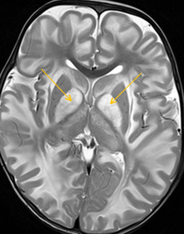
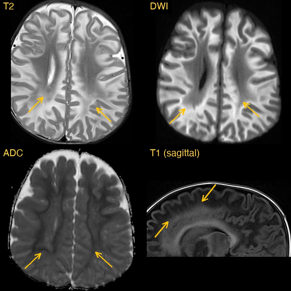
The white matter just under the cortex, including the U-fibres, was bright on T2 and FLAIR in every lobe of both hemispheres. The white matter deeper in, around the ventricles, and the corpus callosum and internal capsules looked normally myelinated. So the disease ran from the outside in. Both globi pallidi, deep grey nuclei, were also bright, while the caudate nuclei and putamina beside them were spared.
The spectrum
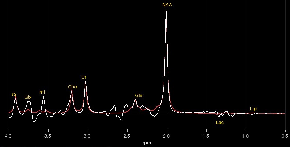
MR spectroscopy measures chemicals inside a small box of brain tissue. Three spectra were taken: two from the right hemisphere white matter (centrum semiovale) at two echo times, and one from the right thalamus and deep grey matter.
Why the spectrum settles it
Widespread abnormal white matter in a young child has a long list of causes, and many of them look alike on the images alone. Spectroscopy can separate them. In most white matter diseases NAA, a marker of healthy nerve cells, goes down. A markedly raised NAA is the classic signature of Canavan disease.
How Monitraq's AI read the scan
- It described the white matter pattern: diffuse, symmetric T2/FLAIR brightness of the subcortical white matter including the U-fibres in the frontal, parietal, temporal and occipital lobes, with the deep white matter, internal capsules and corpus callosum myelinated, giving a centripetal, outside-in pattern.
- It checked the diffusion: the abnormal white matter was bright on DWI with ADC not raised, equal to or below the cortex, so diffusion was restricted.
- It looked at the deep grey matter: both globi pallidi were symmetrically bright on T2 and DWI, the thalami were mildly heterogeneous, and the caudate nuclei and putamina were spared.
- It read the spectra: all three were of good quality. NAA was markedly raised and choline low at both sites, white matter and deep grey matter, at both echo times, with no lactate.
- It put the images and the chemistry together: subcortical white matter disease with restricted diffusion, pallidal involvement and markedly raised NAA made Canavan disease the leading diagnosis. It recommended urine N-acetylaspartic acid and ASPA gene testing to confirm it, radiologist review of the raw spectra, and a metabolic and genetics referral.
Dr Priti, the radiologist who reviewed this case, reported Canavan disease.
What is Canavan disease?
Canavan disease is a rare inherited leukodystrophy, a disease of the brain's white matter. It is autosomal recessive and caused by changes in the ASPA gene, which makes the enzyme aspartoacylase. Without it, N-acetylaspartic acid (NAA) builds up in the brain and the white matter becomes waterlogged and spongy. The typical form begins in infancy, with an enlarging head, floppy muscles and delayed development.
MRI shows diffuse white matter change that usually starts under the cortex, often with the globi pallidi and thalami involved. The raised NAA peak on spectroscopy is the strongest imaging clue. The diagnosis is confirmed by high NAA in the urine and by genetic testing.
The Monitraq draft report
Patient details removed. Shown as generated, before radiologist sign-off.
Technique
3 T brain MRI: axial T2 TSE (4 mm), sagittal T1 TSE-IR (4 mm), coronal T2 FLAIR (4 mm), axial DWI trace with ADC map (4 mm), axial SWI magnitude (2 mm), axial T1 FL2D fat-saturated (4 mm), T1 localiser reformats, and single-voxel PRESS spectroscopy at TE 30 and TE 135 (right centrum semiovale) and TE 30 (right thalamus/deep grey matter).
Findings
White matter / deep grey matter: subcortical white matter of all lobes bilaterally, globi pallidi, thalami
Diffuse, symmetric T2/FLAIR hyperintensity of the subcortical white matter including the U-fibres in frontal, parietal, temporal and occipital lobes, dark on T1 and bright on DWI with ADC not raised (equal to or below cortex), i.e. restricted diffusion; the deep periventricular white matter, internal capsules and corpus callosum are T2 dark and T1 bright (myelinated), giving a centripetal, outside-in pattern. Both globi pallidi are symmetrically T2 bright and DWI bright, with mildly heterogeneous thalamic signal, while the caudate nuclei and putamina are spared. No mass effect, cysts, haemorrhage or SWI blooming; ventricles are not enlarged.
MR spectroscopy: right centrum semiovale/parietal white matter and right thalamus/deep grey matter
All three spectra are of good quality (linewidth 3-5.8 Hz, SNR 108-399). White matter voxel: NAA/Cr 3.77 at TE 30 and 3.23 at TE 135 (markedly raised), Cho/Cr 0.80 and 0.75 (low), mI/Cr 2.46 (raised), no lactate; lipid/macromolecule signal at 0.9 and 1.3 ppm at short TE only, absent at TE 135; Glx/Cr 5.54 and possible succinate at 2.4 ppm are likely fit artefacts from NAA and Glx multiplets. Deep grey voxel: NAA/Cr 2.65 (raised), Cho/Cr 0.45 (low), mI/Cr 1.18, no lactate; overall pattern of markedly raised NAA with reduced choline at both sites.
Impression
- Diffuse symmetric subcortical (U-fibre) white matter T2/FLAIR hyperintensity with restricted diffusion, bilateral globus pallidus and probable thalamic involvement, sparing of the caudate and putamina, and markedly raised NAA with low choline on spectroscopy: Canavan disease is the leading diagnosis.
- Alexander disease is the main alternative (frontal predominance, periventricular rim and enhancement would favour it); MLC, L-2-hydroxyglutaric aciduria and hypomyelination are less likely given the raised NAA and absence of cysts.
- Myelination of the subcortical white matter is abnormal for the patient's age and does not reflect simple maturational delay; central structures (internal capsules, corpus callosum, deep white matter) are myelinated.
- No hydrocephalus, haemorrhage, mass or malformation.
Recommendations
- Urine N-acetylaspartic acid (organic acids) and ASPA gene testing to confirm Canavan disease.
- Radiologist review of the raw spectra, which were machine-processed.
- Clinical correlation with head circumference (macrocephaly), developmental history and hypotonia.
- Clarify whether gadolinium was given on the axial T1 fat-saturated series; if Alexander disease remains in question, post-contrast T1 and GFAP testing.
- Metabolic/genetics referral.
A draft for the radiologist
A good radiologist reads the images and the chemistry together: this white matter pattern, and this NAA peak. Monitraq's AI is built to do the same, and to show its reasoning so the radiologist can check it quickly.
It gives a draft, not a diagnosis. The AI flags, describes and suggests. A radiologist confirms, corrects and signs.
Monitraq's AI produces draft reports to support qualified radiologists. It is not a diagnosis and is not intended for use by patients. All case details have been removed or generalised to protect patient privacy, and the images have been cropped and stripped of identifying information.