Inverted papilloma on MRI: how Monitraq's AI read a sinonasal mass
This case shows how an inverted papilloma looks on MRI: a lobulated mass filling the nose, with a folded, brain-like pattern after contrast. Here is how Monitraq's AI described it, what it favoured, and the one safety step it put before any biopsy.
MRI findings
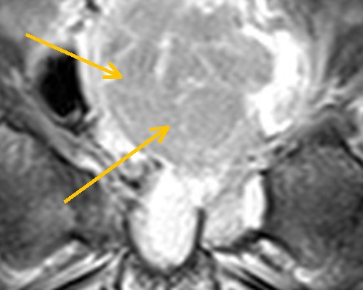
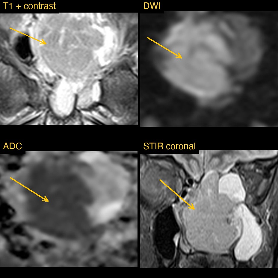
A patient had an MRI of the head that included the sinuses. A large, lobulated solid mass filled and expanded the nasal cavity. It sat mainly on the left. It reached up to the roof of the ethmoid sinuses and the cribriform region, and back through the opening at the back of the nose into the nasopharynx.
Beside the mass, the left maxillary sinus and left ethmoid cells were full of fluid. The right maxillary sinus still held air. The brain itself looked normal, with no bleed, swelling or abnormal enhancement.
How Monitraq's AI read the scan
- It described the mass: a lobulated solid mass expanding the nasal cavity, centred on the left, intermediate on STIR and similar to muscle on T1.
- It separated tumour from fluid: the mass was distinct from the obstructed secretions beside it. Those secretions were very bright on STIR, bright on ADC and did not enhance. The mass was bright on DWI with intermediate to mildly low ADC, below that of the fluid.
- It named the key sign: after contrast the mass enhanced heterogeneously with a convoluted, striated, cerebriform internal pattern.
- It gave a ranked differential: the appearance favoured inverted papilloma. Malignancy, including papilloma-associated squamous cell carcinoma and olfactory neuroblastoma, was the main alternative, with a polyp less likely.
- It flagged a safety point: the mass reached the cribriform plate and ethmoid roof, and an intact bony barrier could not be confirmed on these images. A basal meningoencephalocele had to be excluded first, so it advised no biopsy until thin-section CT of the skull base had been done.
- It listed next steps: thin-section CT with coronal reconstructions of the skull base and sinuses, thin coronal heavily T2-weighted and post-contrast images through the anterior skull base, and ENT and skull-base team review.
Dr Priti, the radiologist who reviewed this case, reported an inverted papilloma.
What is inverted papilloma?
Inverted papilloma is a benign tumour of the lining of the nose and sinuses. Its name comes from the way the surface cells grow inwards, into the underlying tissue, instead of outwards. It usually arises from the lateral wall of the nasal cavity and is usually on one side only.
Although benign, it matters for three reasons. It can grow into nearby sinuses and press on the orbit or skull base. It tends to come back if it is not removed completely. And a minority are linked with squamous cell carcinoma, either at diagnosis or later. Imaging helps to map its extent and to look for features that raise concern for cancer.
Key MRI signs of inverted papilloma
- A unilateral, lobulated mass in the nasal cavity, often reaching into the maxillary sinus or ethmoid cells.
- A convoluted cerebriform pattern: alternating bands that look like folded brain gyri on T2 and on T1 after contrast.
- Blocked secretions in nearby sinuses that are brighter on T2 and do not enhance, which helps to show the true size of the tumour.
- On CT, focal thickening of bone (hyperostosis) often marks the site where the tumour is attached.
- Loss of the cerebriform pattern, bone destruction or spread beyond the sinuses should raise concern for an associated cancer.
Differential diagnosis
The AI's own list was inverted papilloma first, then malignancy (papilloma-associated squamous cell carcinoma or olfactory neuroblastoma), then polyp. In practice a nasal polyp is often bilateral and does not show the striated pattern. Olfactory neuroblastoma arises high in the nose near the cribriform plate and can spread into the skull. When a mass reaches the skull base, a meningoencephalocele, brain tissue herniating through a bony defect, must be ruled out before anyone takes a sample.
Frequently asked questions
What is the cerebriform pattern in inverted papilloma?
It is a series of alternating bright and dark bands inside the mass on T2 and post-contrast T1 images that resemble the folds of the brain. It is considered a characteristic sign of inverted papilloma, though it is not seen in every case.
Why is MRI used as well as CT for a sinonasal mass?
CT shows the bone best, including hyperostosis and erosion. MRI separates tumour from trapped secretions and shows spread into the orbit, skull base or brain, which CT can struggle to do.
Can inverted papilloma turn into cancer?
A minority of inverted papillomas are associated with squamous cell carcinoma. Features such as bone destruction or loss of the usual internal pattern should prompt careful assessment.
A draft for the radiologist
This case shows two things a good report does. It names the most likely diagnosis from a specific sign, and it puts a safety step before an invasive procedure. Here, the AI said the biopsy should wait until CT had checked the skull base.
It gives a draft, not a diagnosis. The AI flags, describes and suggests. A radiologist confirms, corrects and signs.
Monitraq's AI produces draft reports to support qualified radiologists. It is not a diagnosis and is not intended for use by patients. All case details have been removed or generalised to protect patient privacy, and the images have been cropped and stripped of identifying information.