One small bright spot deep in the brain: how Monitraq's AI reads an acute lacunar stroke
A brain MRI showed a single small bright spot deep in the left side of the brain. Small as it was, it sat in one of the most tightly packed pathways in the brain. Here is how Monitraq's AI worked through the scan.
The scan
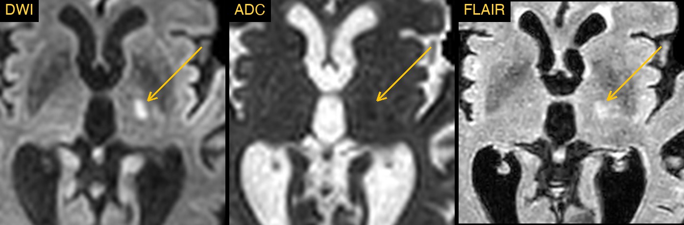
This brain MRI comes from a public research collection of stroke scans. It included diffusion-weighted imaging (DWI), the sequence that lights up brain tissue whose cells have recently been injured, and the matching ADC map, which helps show whether that brightness is a true sign of fresh injury.
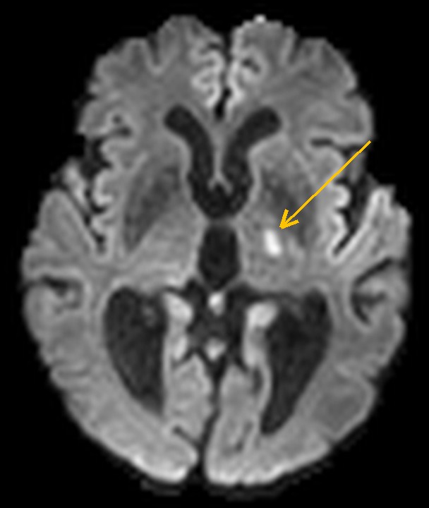
Deep in the left side of the brain, where the thalamus meets the internal capsule, there was one small, oval bright spot on DWI.
Why a small spot matters
A lesion of about a centimetre is easy to pass over on a busy scan. But the posterior limb of the internal capsule carries the motor fibres to the opposite side of the body in a very small space, so a small stroke here can matter far more than its size suggests. And in a stroke the time window for treatment and work-up is short, so a finding like this needs attention the same day.
How Monitraq's AI read the scan
- It found and described the spot: a focal, ovoid DWI-bright lesion of about 8 to 10 mm in the left posterolateral thalamus and the posterior limb of the internal capsule, seen on five neighbouring slices.
- It checked the ADC map: on the matching ADC slices the spot was subtly dark compared with the same place on the other side. Bright on DWI and dark on ADC is the pattern of restricted diffusion, in keeping with an acute infarct.
- It named the artery territory: the spot lay in small-vessel territory, supplied by small penetrating arteries such as the thalamogeniculate, anterior choroidal or lenticulostriate branches. That fits a lacunar infarct, a stroke from one small deep artery.
- It kept the alternative in view: because the ADC change was subtle, it noted that T2 shine-through or partial volume had to be excluded, and asked for the full DWI and ADC series, with contiguous slices and reformats, to be reviewed at that spot.
- It flagged the scan for same-day confirmation and listed the next steps: correlate with the time the symptoms began and, if the infarct is confirmed as acute, proceed with stroke work-up.
The expert lesion segmentation that comes with this public dataset marks an acute left internal capsule and thalamic lacunar infarct.
What is a lacunar infarct?
A lacunar infarct is a small stroke, usually under 15 mm across, deep in the brain. It happens when one of the tiny penetrating arteries that feed the basal ganglia, internal capsule, thalamus or brainstem becomes blocked. High blood pressure and diabetes are the usual background. Depending on where it lands, a lacunar stroke can cause weakness of the face, arm and leg on one side, numbness on one side, or both.
On MRI an acute lacunar infarct is bright on DWI and dark on the ADC map in the first days. Over weeks it can leave a small fluid-filled hole, the lacune that gives the condition its name.
A draft for the radiologist
A good radiologist does not let a small spot slide just because it is small, and checks DWI against ADC before calling it acute. Monitraq's AI is built to do the same, and to show its reasoning so the radiologist can check it quickly.
It gives a draft, not a diagnosis. The AI flags, describes and suggests. A radiologist confirms, corrects and signs.
Monitraq's AI produces draft reports to support qualified radiologists. It is not a diagnosis and is not intended for use by patients. All case details have been removed or generalised to protect patient privacy, and the image has been cropped and stripped of identifying information. Image: ISLES 2022 ischemic stroke lesion segmentation challenge dataset, CC BY 4.0.