Apert syndrome on brain MRI: how Monitraq's AI read it
This brain MRI belongs to a child with Apert syndrome, a genetic form of craniosynostosis. The skull shape, the ventricles and the back of the posterior fossa all carried clues. Here is how Monitraq's AI worked through the scan.
MRI findings
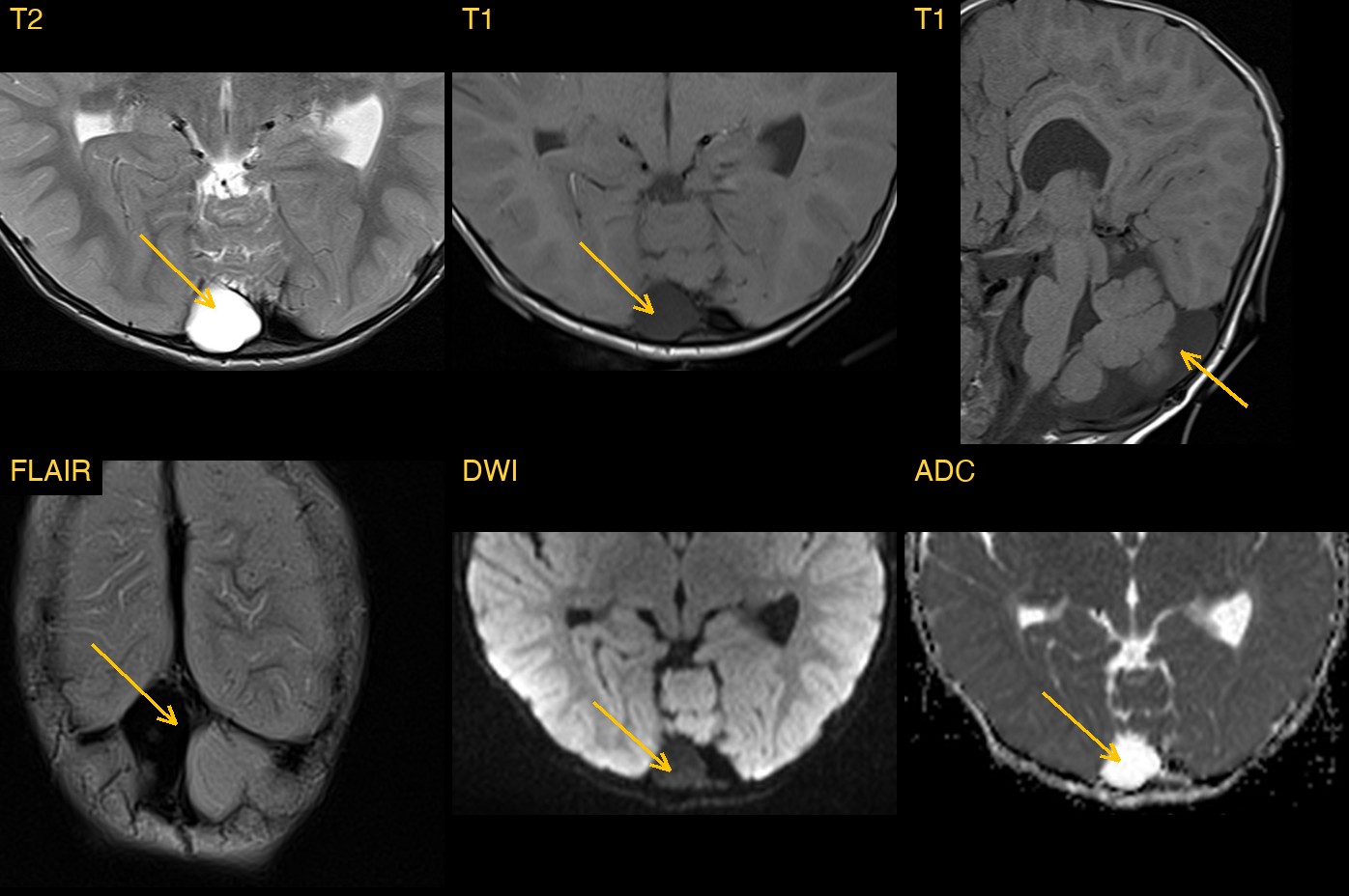
The study had T2, coronal FLAIR, susceptibility-weighted (SWI), diffusion-weighted (DWI) and ADC images, axial and sagittal T1, and a fat-suppressed coronal T2. There was no contrast, and no clinical history was given.
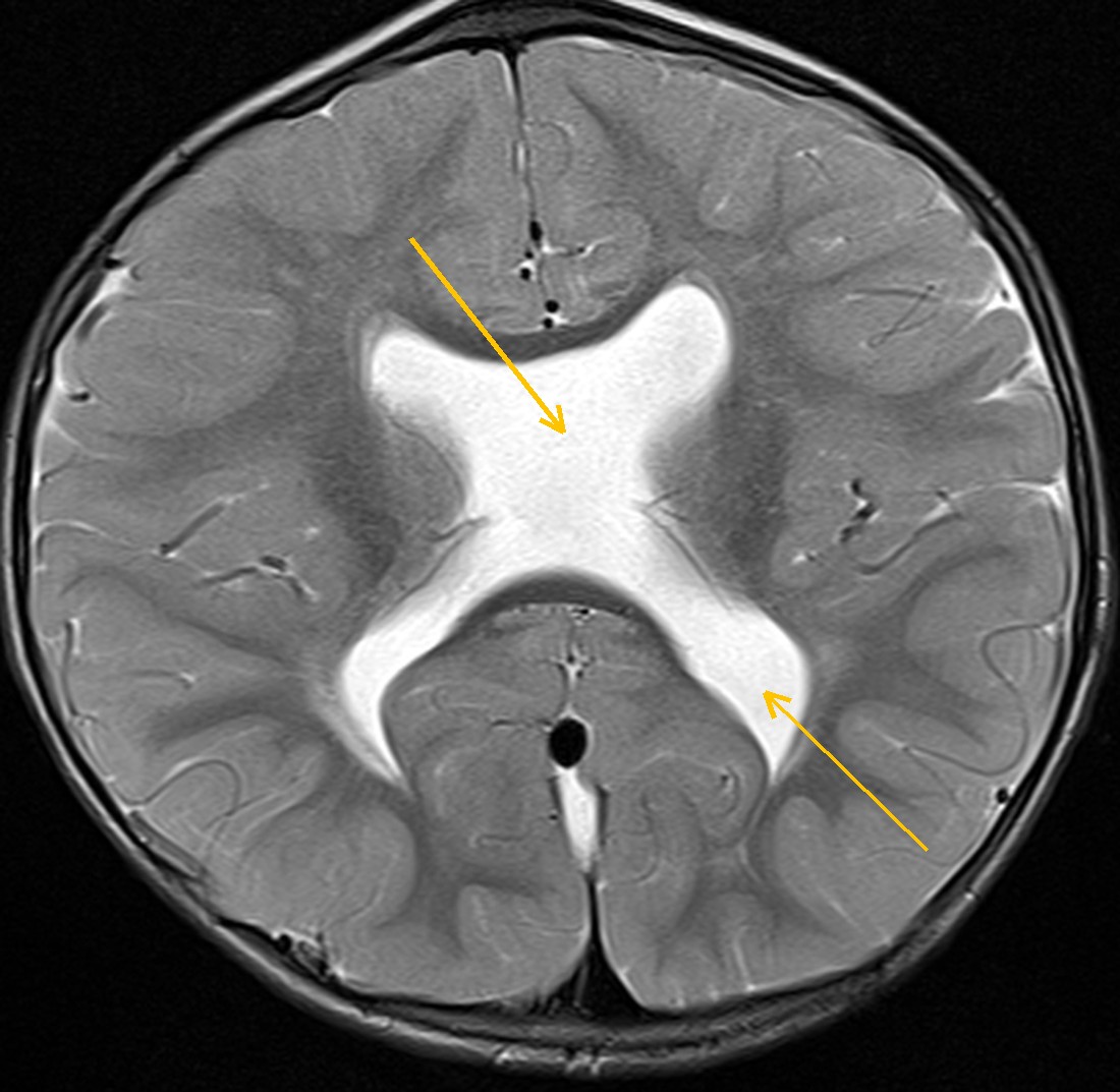
The first clue is the outline of the skull. On axial images it is almost as wide as it is long, and on the midline sagittal image it is taller than it is long.
Inside, the septum pellucidum, the thin sheet that normally separates the two frontal horns, is missing. The frontal horns form a single cavity, and the lateral ventricles are enlarged. Lower down, a large pocket of CSF sits behind the cerebellum.
How Monitraq's AI read the scan
- It described the skull shape: a short, wide and tall skull with a steep forehead and a peaked vertex on coronal images, a turribrachycephalic shape. It measured a cephalic index of about 0.95, above the usual range of about 0.75 to 0.85. It added that the eyes looked prominent relative to the outer orbital rims, suggesting shallow orbits.
- It described the ventricles: the septum pellucidum is absent, and the frontal horns form a single cavity with a flat roof and a downward-pointing contour on coronal images. The lateral ventricles are enlarged, with an Evans index of about 0.37. The third and fourth ventricles are normal in size, there is no fluid seeping into the surrounding white matter, the corpus callosum is present and the gap between the hemispheres is complete.
- It characterised the posterior fossa: a large retrocerebellar CSF space, about 20 x 30 mm on the sagittal image, following CSF on every sequence. The vermis and fourth ventricle look normal and the cerebellum is not compressed. It favoured a mega cisterna magna, with a retrocerebellar arachnoid cyst as the alternative.
- It put the findings together: a turribrachycephalic skull with prominent eyes, absent septum pellucidum and enlarged lateral ventricles, suspicious of syndromic coronal craniosynostosis (Crouzon, Apert or Pfeiffer), with septo-optic dysplasia as the alternative explanation for the absent septum.
- It listed next steps: low-dose 3D CT of the skull to assess the coronal and other sutures; craniofacial and genetic assessment, including FGFR2 and FGFR3 testing and examination of the hands and feet for fused or broad digits; eye and hormone checks because of the absent septum; thin-section imaging of the pituitary and optic pathways; and follow-up of head circumference and signs of raised pressure.
It was also clear that MRI cannot show whether the skull sutures are open or fused. That needs CT.
Dr Priti, the radiologist who reviewed this case, reported Apert syndrome: syndromic craniosynostosis (turribrachycephaly, exorbitism, midface hypoplasia) with absent septum pellucidum, ventriculomegaly and a retrocerebellar CSF collection.
What is Apert syndrome?
Apert syndrome is a genetic condition caused by changes in the FGFR2 gene. It is one of the syndromic craniosynostoses, in which skull sutures fuse too early. In Apert syndrome the coronal sutures are typically fused on both sides, so the skull cannot grow front to back. It grows upwards and sideways instead, giving the short, tall head called turribrachycephaly.
The midface is underdeveloped and the orbits are shallow, so the eyes look prominent. The feature that sets Apert apart from similar syndromes is fusion of the fingers and toes (syndactyly). Brain differences such as large ventricles and anomalies of the septum pellucidum and corpus callosum are also described.
Care is led by a craniofacial team. Brain MRI helps by showing the ventricles, the midline structures and signs of raised pressure.
Key MRI signs of Apert syndrome
- A short, wide and tall skull (turribrachycephaly) from early fusion of both coronal sutures.
- Shallow orbits with prominent eyes (exorbitism) and an underdeveloped midface.
- Enlarged lateral ventricles.
- Absent or abnormal septum pellucidum, and sometimes corpus callosum anomalies.
- Fused sutures are confirmed on CT, not MRI.
Differential diagnosis
- Crouzon syndrome. Similar skull and face, but the hands and feet are normal.
- Pfeiffer syndrome. Similar skull shape, with broad thumbs and big toes.
- Non-syndromic bicoronal synostosis. Fused coronal sutures without facial or limb features.
- Positional brachycephaly. A flat back of the head with open sutures and normal orbits.
- Septo-optic dysplasia, for the absent septum: look for small optic nerves and chiasm and pituitary anomalies.
Frequently asked questions
Can MRI diagnose craniosynostosis?
MRI shows the skull shape and the brain well, but not whether the sutures are fused. Low-dose CT with 3D reconstruction is the usual test for the sutures.
How is Apert syndrome told apart from Crouzon syndrome?
Both have similar skull and face changes. Apert syndrome has fusion of the fingers and toes, while Crouzon syndrome does not. Genetic testing confirms the diagnosis.
How does a mega cisterna magna differ from a retrocerebellar arachnoid cyst?
A mega cisterna magna is an enlarged CSF space with a normal vermis and fourth ventricle and no mass effect. An arachnoid cyst may press on the vermis or cerebellum; both it and a mega cisterna magna can scallop the occipital bone. Both follow CSF signal on MRI.
A draft for the radiologist
In this scan no single finding gave the answer. The AI linked the skull shape, the eyes, the absent septum and the large ventricles into one pattern, and said which tests would confirm it.
It gives a draft, not a diagnosis. The AI flags, describes and suggests. A radiologist confirms, corrects and signs.
Monitraq's AI produces draft reports to support qualified radiologists. It is not a diagnosis and is not intended for use by patients. All case details have been removed or generalised to protect patient privacy, and the images have been cropped and stripped of identifying information.