Bright tracts in a newborn: how Monitraq's AI read a pattern of maple syrup urine disease
A newborn baby had a brain MRI. On diffusion imaging, the brain pathways that are already myelinated at birth lit up on both sides, from the brainstem to the internal capsules. Spectroscopy showed an unusual signal at 0.9 ppm. Here is how Monitraq's AI put the two together.
The scan
The study included T2, FLAIR, T1, diffusion-weighted imaging (DWI) with its ADC map, susceptibility imaging (SWI) and two MR spectra. No clinical history came with it.
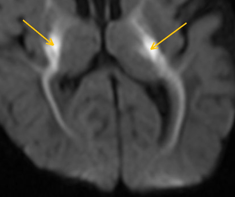
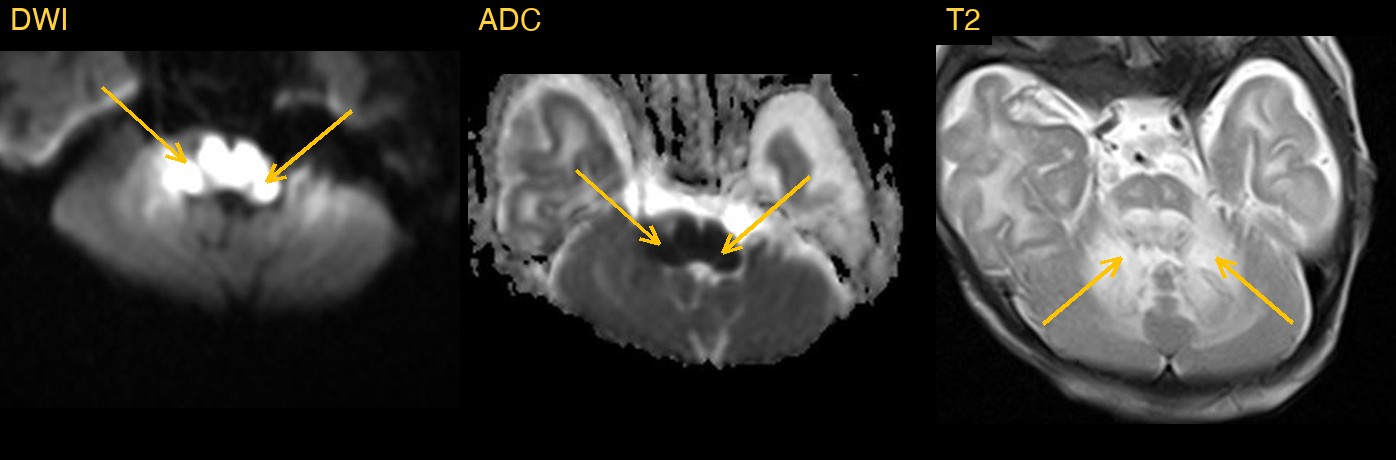
The striking finding was on DWI. A newborn brain is mostly unmyelinated, but a few pathways already carry myelin at birth: the back of the brainstem, the white matter of the cerebellum, the cerebral peduncles, the posterior limbs of the internal capsules and the motor tracts that run up to the region around the central sulcus. In this baby, exactly those pathways were very bright on DWI and dark on the ADC map, on both sides, while the unmyelinated white matter and the cortex were spared.
Why the distribution matters
Restricted diffusion in a newborn often brings oxygen deprivation around birth to mind first. That kind of injury, however, tends to involve the putamina, the cortex around the central sulcus and the hippocampi. Here the change followed the myelinated tracts instead, with marked swelling of the back of the brainstem and the cerebellar white matter. A pattern that tracks myelin, rather than blood supply, points towards a metabolic cause.
What the spectroscopy added
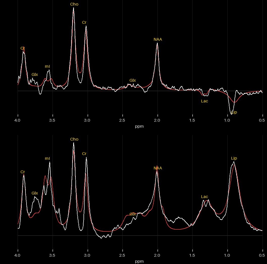
Two single-voxel spectra were acquired from the centre of the brain, one at a long echo time and one at a short echo time. Spectroscopy measures the chemical make-up of a small block of tissue. Each peak sits at a fixed position on the scale, measured in parts per million (ppm).
How Monitraq's AI read the scan
- It traced the pattern: marked symmetric DWI brightness with matching ADC darkness, following the tracts that are normally myelinated at this age. It ran from the dorsal medulla and pons and the cerebellar white matter around the dentate nuclei, through the cerebral peduncles and the posterior limbs of the internal capsules with the ventrolateral thalami and pallidi, up the corticospinal tracts to the perirolandic white matter.
- It checked the other sequences: on T2 the dorsal brainstem and cerebellar white matter were swollen and abnormally bright where they should be dark, with matching FLAIR brightness along the brainstem tracts. The fourth ventricle stayed open, and there was no hydrocephalus, haemorrhage or mass.
- It read the spectra: a 0.9 ppm peak, inverted at the long echo time and large and upright at the short one, together with lactate and reduced NAA. In this setting it read the 0.9 ppm signal as branched-chain amino and keto acids, not an abscess, because there was no cavity.
- It named the diagnosis and the alternatives: the pattern of maple syrup urine disease in metabolic crisis. Non-ketotic hyperglycinaemia was the main alternative, kept open by a small 3.55 ppm peak that could be glycine or myo-inositol. Hypoxic-ischaemic injury was judged less likely.
- It marked the scan urgent and asked for same-day radiologist review and direct contact with the neonatal team. It listed the tests that would confirm the diagnosis: plasma amino acids (leucine, isoleucine, valine and alloisoleucine), urine organic acids and ketoacids, blood gas, glucose, ammonia and lactate, and paired CSF and plasma glycine. It also advised early involvement of the metabolic team and a follow-up MRI with DWI and spectroscopy after treatment.
Dr Priti, the radiologist who reviewed this case, reported maple syrup urine disease.
What is maple syrup urine disease?
Maple syrup urine disease (MSUD) is a rare inherited disorder in which the body cannot break down three amino acids: leucine, isoleucine and valine, known as the branched-chain amino acids. They and their keto acids build up in the blood and are toxic to the brain. The name comes from the sweet smell of the urine.
In the classic form, a baby who seems well at birth becomes unwell within the first days of life, with poor feeding, drowsiness and, without treatment, worsening encephalopathy. On MRI the build-up causes marked swelling with restricted diffusion in the parts of the brain that are already myelinated, and spectroscopy can show a peak at about 0.9 ppm from the branched-chain amino and keto acids. The diagnosis is confirmed with blood and urine tests. It is a medical emergency: treatment lowers the toxic levels quickly and then limits these amino acids in the diet for life.
The Monitraq draft report
Patient details removed. Shown as generated, before radiologist sign-off.
Technique
3 T brain MRI: axial T2 TSE, sagittal T1 TSE-IR, coronal T2 FLAIR, axial DWI trace with ADC map, axial SWI (magnitude and processed), axial T1 TIR, and two single-voxel spectra (TE 135 ms and TE 30 ms, TR 2000 ms). No contrast series.
Findings
Myelinated tracts. Marked symmetric DWI brightness with matching ADC darkness follows the tracts that are normally myelinated at this age. It runs from the dorsal medulla and pons and the cerebellar white matter around the dentate nuclei, through the cerebral peduncles and posterior limbs of the internal capsules with the ventrolateral thalami and pallidi, up the corticospinal tracts to the perirolandic white matter. On T2 the dorsal brainstem and cerebellar white matter are swollen and abnormally bright where they should be dark, with corresponding FLAIR brightness along the brainstem tracts; the fourth ventricle stays open and there is no hydrocephalus or haemorrhage.
MR spectroscopy. MRS1 (TE 135, good quality) shows NAA/Cr about 0.78, Cho/Cr about 1.33 and Cho/NAA about 1.70, with an inverted lactate doublet at 1.33 ppm and a distinct inverted signal at 0.9 ppm; in this context that 0.9 ppm signal fits branched-chain amino and keto acids, not an abscess (there is no cavity), and the small 3.55 ppm peak may be glycine or residual myo-inositol. MRS2 (TE 30, fair quality, residual fit structure) shows a large upright 0.9 ppm peak (/Cr about 3.06), a 1.3 ppm peak that overlaps lactate and lipid, NAA/Cr about 1.08, Cho/Cr about 1.41 and mI/Cr about 1.98 (high mI is usual for a neonate). Acetate and succinate are absent.
Impression
- Symmetric restricted diffusion and swelling of the myelinated tracts (dorsal brainstem, cerebellar white matter, cerebral peduncles, posterior limbs of the internal capsules, ventrolateral thalami and pallidi, perirolandic corticospinal tracts) in a neonate: the pattern of maple syrup urine disease in metabolic crisis. Non-ketotic hyperglycinaemia is the main alternative, and hypoxic-ischaemic injury is less likely.
- Spectroscopy supports this: a 0.9 ppm peak (branched-chain amino/keto acids) with lactate and reduced NAA. A possible glycine peak keeps NKH in the differential. Both spectra are machine-processed and need review.
- No haemorrhage, hydrocephalus or mass; brain development otherwise appropriate for age.
Recommendations
- Same-day radiologist review and direct communication to the neonatal team for urgent metabolic work-up: plasma amino acids (leucine, isoleucine, valine, alloisoleucine), urine organic acids and ketoacids, blood gas, glucose, ammonia and lactate, and paired CSF/plasma glycine.
- Early involvement of the metabolic specialist team.
- Review the raw spectra; if NKH remains in question, a long-TE (TE 144-288) spectrum would separate glycine from myo-inositol.
- Clinical correlation with the perinatal history to weigh hypoxic-ischaemic injury.
- Follow-up MRI with DWI and MRS after treatment to document resolution.
Two incidental items listed after the main findings (a susceptibility artefact and a normal-variant cavum septum pellucidum) are not shown.
A draft for the radiologist
In a newborn, the pattern on the images can be the first clue to a metabolic emergency, and the next steps are laboratory tests that need to happen quickly. Monitraq's AI is built to read the images and the spectra together, to say what the pattern points to, and to show its reasoning so the radiologist can check it quickly.
It gives a draft, not a diagnosis. The AI flags, describes and suggests. A radiologist confirms, corrects and signs.
Monitraq's AI produces draft reports to support qualified radiologists. It is not a diagnosis and is not intended for use by patients. All case details have been removed or generalised to protect patient privacy. The images have been cropped and stripped of identifying information, and the draft report is shown with patient details and image references removed.