Mucormycosis sinusitis with cerebritis on MRI: how Monitraq's AI read it
This brain MRI shows mucormycosis: infection filling most of the paranasal sinuses, with inflamed, swollen frontal lobes above them. Here is how Monitraq's AI worked through the scan and why it put an invasive fungal infection first.
MRI findings
The study had T2, FLAIR, diffusion-weighted (DWI) and ADC images, susceptibility-weighted images (SWI) and fat-suppressed T1 images after contrast in three planes. No clinical history was given.
Two things stand out. First, nearly every sinus is diseased. The frontal, ethmoid and maxillary sinuses are filled with fluid and thickened lining, more on the right maxillary side, and the sphenoid lining is thickened too. After contrast, parts of the nasal and ethmoid lining do not enhance at all.
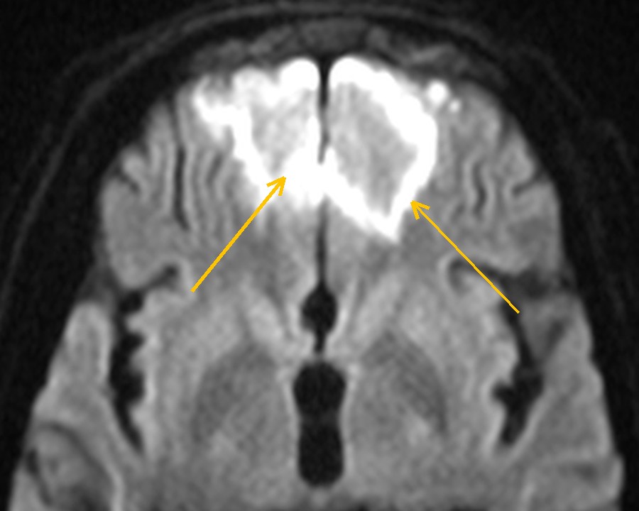
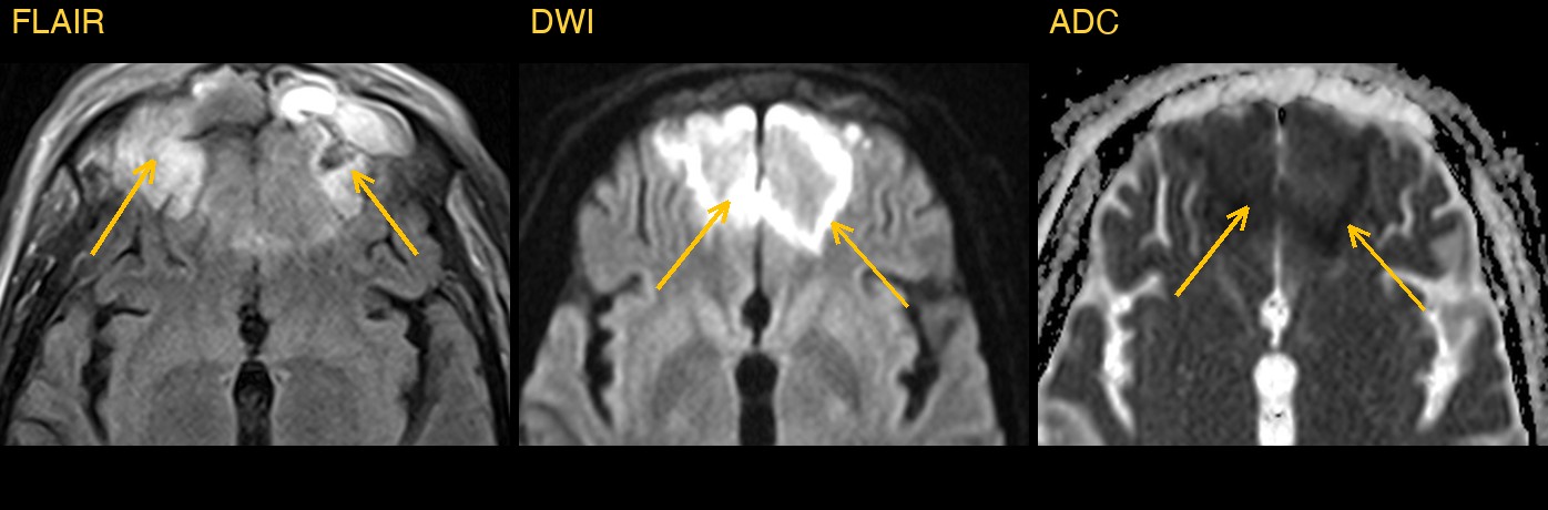
Second, the frontal lobes just above the sinuses are abnormal on both sides. The cortex and the white matter beneath it are swollen and bright on FLAIR, and parts of the cortex are bright on DWI and dark on ADC.
How Monitraq's AI read the scan
- It mapped the sinus disease: pansinus involvement, with the frontal, ethmoid and maxillary sinuses opacified by T2/FLAIR-bright secretions and thickened mucosa, right maxillary more than left, and mucosal thickening in the sphenoid sinus.
- It looked at how the lining enhanced: heterogeneous enhancement of the nasal and ethmoid mucosa, with areas of non-enhancing mucosa. It also raised possible stranding of the fat behind the right maxillary sinus, in the right pterygopalatine and masticator regions and in the right facial soft tissues.
- It found a collection in front of the brain: a bifrontal extra-axial collection, most likely epidural and crossing the midline, about 7 mm thick, with restricted diffusion and a thick enhancing rim: an empyema, which it flagged as urgent.
- It followed the infection upwards: ill-defined T2/FLAIR brightness in the orbitofrontal and frontopolar cortex and the white matter beneath it on both sides, with patchy cortical diffusion restriction and effaced sulci, and no discrete ring-enhancing abscess. It read this as frontal cerebritis on both sides, secondary to the sinusitis.
- It named the likely cause: invasive fungal sinusitis (mucormycosis or aspergillosis) as the leading cause, with complicated bacterial sinusitis as the alternative. The pointers it gave for fungal infection were non-enhancing (necrotic) mucosa, infiltration of the fat beyond the sinus, and a diabetic or immunocompromised patient. It added that biopsy would confirm it.
- It flagged the scan as urgent and listed next steps: urgent ENT and neurosurgical review for endoscopic sinus evaluation with biopsy and fungal staining; blood glucose, HbA1c and immune status; CT of the sinuses and skull base for bone erosion; MR venography to check the superior sagittal and cavernous sinuses for septic thrombosis; and an early follow-up MRI with contrast to watch for abscess formation.
It also said what the study could not show: without fat-suppressed orbital images or a dedicated coronal T2, subtle spread to the orbit, orbital apex or cavernous sinus could not be excluded.
Dr Priti, the radiologist who reviewed this case, reported pansinusitis due to mucormycosis with cerebritis.
What is mucormycosis?
Mucormycosis is a fast-moving fungal infection caused by moulds of the order Mucorales, such as Rhizopus and Mucor. These moulds are common in the environment and rarely harm healthy people. The infection is seen mainly in people with poorly controlled diabetes, especially with ketoacidosis, and in people whose immunity is weakened by treatment or illness.
The rhino-orbito-cerebral form starts in the nose and sinuses. The fungus invades blood vessels, which causes tissue death, and it can spread into the orbit, the cavernous sinus and the brain, sometimes before the bone looks destroyed.
It is a medical emergency. Treatment usually combines urgent surgical removal of dead tissue with antifungal drugs and correction of the underlying problem, so early recognition on imaging matters.
Key MRI signs of mucormycosis
- Thickened or opacified sinus lining, often asymmetric.
- Non-enhancing, dead mucosa after contrast, including the "black turbinate" sign in the nasal cavity.
- Infiltration of the fat outside the sinus walls, such as behind the maxillary sinus or in the pterygopalatine fossa, even when the bone looks intact.
- Spread into the orbit, orbital apex or cavernous sinus.
- Intracranial spread: cerebritis or abscess, and vessel involvement causing infarcts.
Differential diagnosis
- Invasive aspergillosis. Also an angioinvasive fungal infection in immunocompromised people. Imaging alone often cannot separate it from mucormycosis, so tissue diagnosis is needed.
- Complicated bacterial sinusitis. Can also spread to the brain. The lining usually enhances evenly, and the fat outside the sinus is usually spared.
- Sinonasal tumour. A solid, enhancing mass with bone destruction, separate from trapped secretions, points to a tumour.
- Other causes of frontal lobe change, such as infarction or a tumour, are less likely when the brain change sits directly above infected sinuses.
Frequently asked questions
What is the black turbinate sign?
It is a nasal turbinate that does not enhance on post-contrast T1 images, because the tissue has died. In the right setting it is an early clue to invasive fungal sinusitis, especially mucormycosis.
What does cerebritis look like on DWI?
Cerebritis is an early, poorly defined brain infection. It is usually bright on T2 and FLAIR and may show patchy restricted diffusion, bright on DWI and dark on ADC, without the well-formed enhancing rim of an abscess.
Is MRI or CT better for invasive fungal sinusitis?
They answer different questions. CT shows bone erosion of the sinus walls and skull base well. Contrast-enhanced MRI is better for non-enhancing mucosa and for spread into the orbit, cavernous sinus and brain.
A draft for the radiologist
Sinus disease is common on brain MRI and is usually trivial. Here the AI treated it as the source of a brain infection, linked the two, and named the fungal cause that needs the fastest response.
It gives a draft, not a diagnosis. The AI flags, describes and suggests. A radiologist confirms, corrects and signs.
Monitraq's AI produces draft reports to support qualified radiologists. It is not a diagnosis and is not intended for use by patients. All case details have been removed or generalised to protect patient privacy, and the images have been cropped and stripped of identifying information.