Bright patches under the cortex: how Monitraq's AI reads tuberous sclerosis
A brain MRI showed several bright patches scattered through the cortex of both hemispheres. Their shape and position pointed to one genetic condition: tuberous sclerosis complex. Here is how Monitraq's AI worked through the scan.
The scan
The patient had a brain MRI with the usual set of sequences. There was no bleed, no stroke and nothing pressing on the brain, and the ventricles were not enlarged.
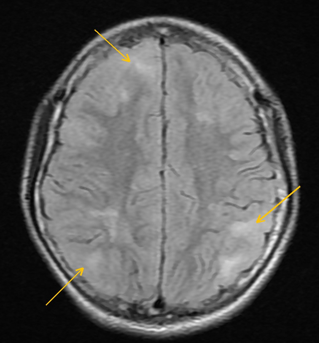
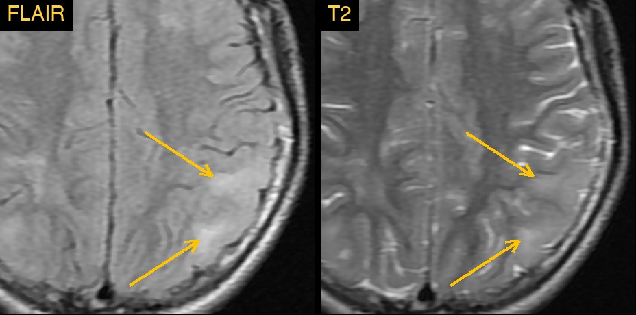
The clue was on FLAIR and T2-weighted images, the sequences that show abnormal water in brain tissue as bright. Several patches in both hemispheres were brighter than the normal cortex around them, more on the left than the right.
Why the shape of the patches matters
Many things can make the cortex look bright on FLAIR: a stroke, inflammation, a tumour or a recent seizure. What separates them is the detail. These patches were wedge-shaped or followed the shape of a gyrus, sat right at the grey-white junction, and made the gyri look fuller rather than thinner. There was no mass effect. That combination is the pattern of a developmental abnormality of the cortex, not of new damage.
How Monitraq's AI read the scan
- It described the patches precisely: several wedge-shaped or gyral areas, bright on T2 and FLAIR and mildly dark on T1, at the grey-white junction and in the subcortical white matter. It placed them in the left frontal, left parietal, left posterior temporal, left paramedian superior frontal, right frontal and right parietal lobes, left more than right.
- It read the pattern: the patches expanded the gyri rather than shrinking them, and blurred the boundary between grey and white matter. They were multifocal and on both sides, but not a mirror image, so they did not fit a symmetric white matter disease. The deep white matter, corpus callosum, deep grey nuclei, brainstem and cerebellum were spared.
- It named the most likely cause: cortical and subcortical tubers of tuberous sclerosis complex. It gave multifocal focal cortical dysplasia as the main alternative.
- It listed the next steps: review the full FLAIR, T2, SWI and post-contrast series to count the tubers; correlate with seizures and other features of tuberous sclerosis in the skin, kidneys, heart and eyes; and consider referral for genetic testing of the TSC1 and TSC2 genes.
Dr Priti, the radiologist who reviewed this case, reported tuberous sclerosis, with the cortex looking swollen and abnormal because of tubers and mild oedema.
What is tuberous sclerosis complex?
Tuberous sclerosis complex is a genetic condition caused by changes in the TSC1 or TSC2 gene. It leads to benign growths in many organs, including the brain, skin, kidneys, heart and eyes.
In the brain, the typical lesions are cortical tubers: patches of cortex that did not form normally during development. Tubers are a common cause of epilepsy in people with the condition. MRI is the main way to see them, and spotting the pattern can lead the clinical team to look for the condition in the other organs it affects.
A draft for the radiologist
A good radiologist does not stop at "bright spots in the cortex". The shape, the position and the effect on the gyri tell the story. Monitraq's AI is built to look at the same details, and to show its reasoning so the radiologist can check it quickly.
It gives a draft, not a diagnosis. The AI flags, describes and suggests. A radiologist confirms, corrects and signs.
Monitraq's AI produces draft reports to support qualified radiologists. It is not a diagnosis and is not intended for use by patients. All case details have been removed or generalised to protect patient privacy, and the image has been cropped and stripped of identifying information.