Carotid space neck mass on MRI: how Monitraq's AI read it
A carotid space mass on neck MRI raises two main questions: is it a paraganglioma or a nerve sheath tumour, and is it safe to biopsy? Here is how Monitraq's AI read a large mass in the left upper neck, and what the radiologist concluded.
MRI findings
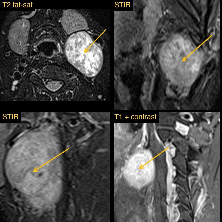
The study was an MRI of the cervical spine and neck, with STIR in the coronal and sagittal planes, axial T2 and T1 Dixon images, and fat-suppressed T1 after contrast. The cervical spine itself was normal.
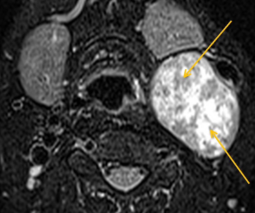
The finding is in the left upper neck, at the angle of the jaw. A single, well-defined oval mass sits in the carotid space, behind the left submandibular gland and along the carotid sheath. It is bright and patchy on fat-suppressed T2 and STIR, and it enhances strongly after contrast.
The mass pushes the left submandibular gland forward, but there is a clear line between the two, and it shows no visible invasion of the muscles around it.
How Monitraq's AI read the scan
- It placed the mass: left upper neck at the angle of the jaw, behind the left submandibular gland, in the carotid space and post-styloid parapharyngeal space at about C2 to C3, measuring about 39 x 44 x 53 mm.
- It described its signal: heterogeneously bright on fat-suppressed T2 and STIR, with a speckled internal pattern and punctate signal voids, dark on the fat-only images, and avidly enhancing after contrast.
- It checked the neighbours: the mass displaced the left submandibular gland forward with a distinct interface, narrowed the left parapharyngeal space, and did not visibly invade the adjacent muscles.
- It gave a ranked differential: carotid body paraganglioma as the leading diagnosis; schwannoma of the vagus nerve or sympathetic chain as the main alternative; and pleomorphic adenoma of the submandibular gland or parotid tail, or a necrotic level II lymph node, as unlikely.
- It set out next steps: CT or MR angiography of the neck to show how the mass relates to the carotid bifurcation; plasma or urine metanephrines; ENT or head and neck surgical referral, avoiding percutaneous biopsy until a paraganglioma has been excluded; and neck ultrasound of the left jugular chain nodes.
It also stated what the study could not show: the relationship of the mass to the carotid bifurcation was only partly covered by these images.
Dr Priti, the radiologist who reviewed this case, reported a schwannoma (neurogenic tumour) of the left carotid space / posterior triangle, separate from the parotid and submandibular glands, bright on T2 and enhancing, with non-enhancing necrotic areas.
The AI's leading diagnosis and the radiologist's diagnosis differ here, and the AI's list carried both. Telling the two apart on imaging can be hard. This is exactly why a radiologist reads and signs every report.
What is a carotid space mass?
The carotid space is a column of tissue on each side of the neck. It holds the carotid artery, the internal jugular vein, the vagus nerve and the sympathetic chain. Masses here are uncommon.
The two most common primary tumours are paragangliomas and schwannomas. Paragangliomas arise from neural crest cells, such as the carotid body at the carotid bifurcation. Schwannomas arise from the sheath of a nerve, most often the vagus nerve or the sympathetic chain. Both usually present as a painless lump.
The distinction changes management. Paragangliomas are very vascular, so biopsy can bleed, and some secrete catecholamines. Schwannomas are less vascular, but surgery can injure the nerve they arise from.
Key MRI signs of a carotid space mass
- A well-defined mass along the carotid sheath, deep to the sternocleidomastoid muscle.
- Displacement of the carotid artery and jugular vein, which helps show where the mass arises.
- Paraganglioma: a "salt-and-pepper" look with flow voids, avid enhancement, and splaying of the internal and external carotid arteries at the bifurcation.
- Schwannoma: bright on T2, often with cystic or non-enhancing areas, and usually without flow voids.
- A fat plane separating the mass from the parotid and submandibular glands.
Differential diagnosis
- Carotid body paraganglioma. Sits at the bifurcation and splays the two carotid branches apart.
- Vagal or sympathetic schwannoma. A vagal schwannoma tends to push the carotid artery and jugular vein apart. A sympathetic schwannoma tends to push both vessels together, without separating them.
- Salivary gland tumour. A pleomorphic adenoma of the parotid tail or submandibular gland is wrapped by a claw of gland tissue.
- Lymph node. A necrotic or metastatic node, often with other abnormal nodes and a primary tumour in the head and neck.
Frequently asked questions
How do you tell a schwannoma from a paraganglioma on MRI?
Paragangliomas usually enhance avidly and show flow voids, giving a salt-and-pepper look. Schwannomas also enhance, often unevenly, often contain cystic or non-enhancing areas, and usually lack prominent flow voids. The way the carotid artery and jugular vein are displaced also helps.
Why avoid biopsy of a carotid space mass?
If the mass is a paraganglioma, it is very vascular and may secrete catecholamines. Imaging and blood or urine tests are used first so that biopsy is not done blindly.
Which MRI sequences are most useful for a neck mass?
Fat-suppressed T2 or STIR show the mass against dark fat, and fat-suppressed T1 after contrast shows how it enhances.
A draft for the radiologist
Here the AI placed the mass, showed it was separate from the submandibular gland, and listed both likely tumour types with the tests that separate them.
It gives a draft, not a diagnosis. The AI flags, describes and suggests. A radiologist confirms, corrects and signs.
Monitraq's AI produces draft reports to support qualified radiologists. It is not a diagnosis and is not intended for use by patients. All case details have been removed or generalised to protect patient privacy, and the images have been cropped and stripped of identifying information.