PRES with venous sinus thrombosis on MRI: how Monitraq's AI read it
This brain MRI shows posterior reversible encephalopathy syndrome (PRES) together with a clot in a dural venous sinus. A teenager's scan had patches of brain swelling on both sides and one venous sinus that looked abnormal. Here is how Monitraq's AI worked through it.
MRI findings
The study had T2, coronal FLAIR, susceptibility-weighted (SWI), diffusion-weighted (DWI) and ADC images, and fat-suppressed T1 images before and after contrast. No history, blood pressure or medication details were given.
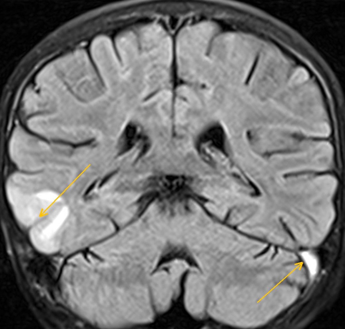
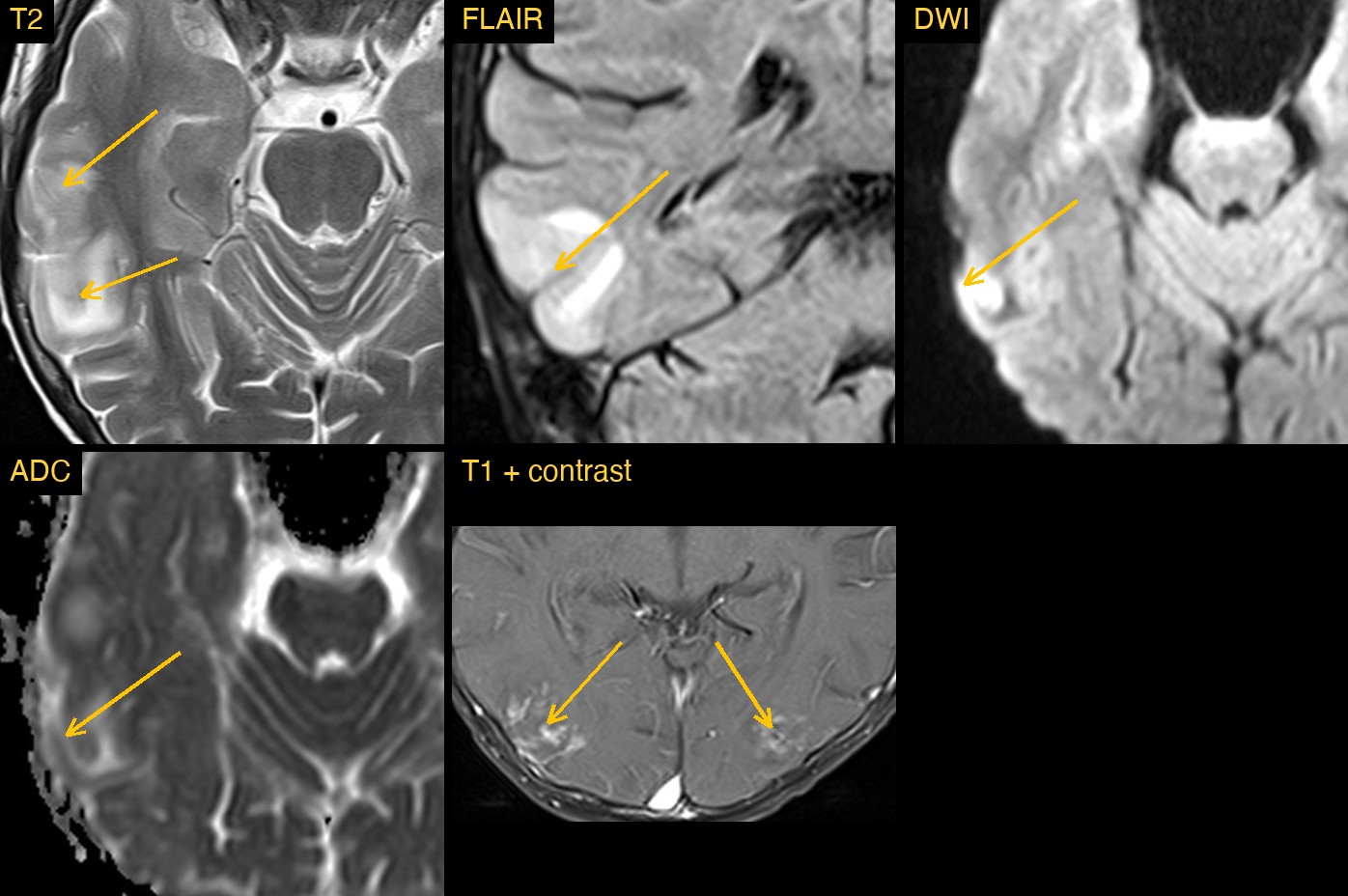
On T2 and FLAIR there were several bright, swollen patches in the cortex and the white matter just beneath it, on both sides of the brain. The largest was in the right posterior temporal and temporo-occipital lobe. Others sat in the left parieto-occipital lobe, the right frontal lobe near the superior frontal sulcus, and both parietal lobes.
The second clue was a vein. The left transverse and sigmoid sinus was bright on FLAIR and on the pre-contrast T1 images, where a normal sinus is dark because blood is flowing through it. The right side looked normal.
How Monitraq's AI read the scan
- It mapped the swelling: multiple cortical and subcortical T2/FLAIR-bright areas with gyral swelling in the right frontal, right posterior temporo-occipital, both parietal and the left parieto-occipital lobes, largest on the right. There was local sulcal effacement but no midline shift or herniation.
- It checked diffusion and blood: no restricted diffusion, with ADC normal or raised, so the swelling was vasogenic oedema. A small right temporal DWI focus had raised ADC and was read as T2 shine-through. On SWI the lesions showed no blooming.
- It looked at contrast: mild patchy gyriform enhancement in the right temporo-occipital, right frontal and left parieto-occipital lesions.
- It named the pattern: multifocal vasogenic oedema suggestive of PRES, with venous oedema from sinus or cortical vein thrombosis and MELAS stroke-like lesions as the main alternatives.
- It did not stop at the brain. It found the left transverse-sigmoid sinus bright on pre-contrast T1 and FLAIR over several sections, while the right kept its normal dark flow void. After contrast the sinuses filled without a clear empty delta sign. It called this suspicious for subacute venous sinus thrombosis, with slow flow as the alternative, and noted no bleeding next to it.
- It listed the next steps: urgent MR or CT venography to confirm or exclude the thrombosis; blood pressure, kidney function and any immunosuppressant, chemotherapy or steroid use as PRES triggers; tests for MELAS and encephalitis if no trigger is found; and a follow-up MRI after treatment to show that the oedema has reversed.
Dr Priti, the radiologist who reviewed this case, reported PRES with dural venous sinus thrombosis.
What is PRES?
Posterior reversible encephalopathy syndrome is brain swelling caused by fluid leaking out of small vessels when the brain can no longer keep its blood flow steady. It is linked to sudden rises in blood pressure, kidney disease, eclampsia and some immunosuppressive and cancer drugs. Patients may have headache, confusion, seizures or visual disturbance.
As the name says, the back of the brain is affected most often, but the frontal lobes, temporal lobes, cerebellum and brainstem can be involved too. The swelling often settles when the cause is treated.
Why venous sinus thrombosis matters here
Cerebral venous sinus thrombosis is a clot in one of the large veins that drain the brain. When drainage is blocked, the brain upstream can swell and bleed. That venous oedema can look very like PRES on T2 and FLAIR, and the two can occur together. A clot also changes treatment, so a sinus that looks bright on T1 or FLAIR should never be passed over. Venography is the usual next test.
Key MRI signs of PRES with venous sinus thrombosis
- Patchy cortical and subcortical T2/FLAIR brightness, often on both sides and most often in the parieto-occipital lobes.
- ADC normal or raised in the swollen areas, showing vasogenic rather than cytotoxic oedema.
- Variable patchy enhancement and, in some patients, small bleeds on SWI.
- Loss of the normal dark flow void in a venous sinus, with bright signal on T1 or FLAIR.
- A filling defect, such as the empty delta sign, after contrast or on venography.
Differential diagnosis
- Venous infarction from sinus or cortical vein thrombosis. Oedema in a venous drainage territory, often with bleeding at the grey-white junction.
- MELAS. A mitochondrial disorder whose stroke-like lesions favour the cortex and do not follow arterial territories. Raised lactate and a lactate peak on MR spectroscopy support it.
- Encephalitis or CNS vasculitis. Usually with fever, abnormal CSF, or infarcts of different ages with restricted diffusion.
- Slow flow in a normal sinus. Can mimic thrombus on routine sequences, which is why venography is needed to confirm it.
Frequently asked questions
How does DWI help tell PRES from a stroke?
In PRES the oedema is vasogenic, so ADC is normal or raised. An acute arterial infarct shows restricted diffusion: bright on DWI and dark on ADC. Some bright spots on DWI in PRES are T2 shine-through, with ADC not dark.
What does venous sinus thrombosis look like on MRI?
The normal dark flow void in the sinus is lost, and the clot may be bright on T1 and FLAIR, depending on its age. After contrast, a filling defect such as the empty delta sign may be seen. MR or CT venography confirms it.
Is PRES always reversible?
Often, but not always. Most patients improve when the cause is treated, but bleeding or infarction can leave lasting damage, so a follow-up MRI is used to check that the oedema has gone.
A draft for the radiologist
Once a pattern as striking as PRES is found, it is easy to stop looking. Here the AI kept going and flagged a venous sinus that did not look normal, the kind of second finding that changes what happens next.
It gives a draft, not a diagnosis. The AI flags, describes and suggests. A radiologist confirms, corrects and signs.
Monitraq's AI produces draft reports to support qualified radiologists. It is not a diagnosis and is not intended for use by patients. All case details have been removed or generalised to protect patient privacy, and the images have been cropped and stripped of identifying information.