Deep brain arteriovenous malformation (AVM) on MRI: how Monitraq's AI read it
This brain MRI shows a deep arteriovenous malformation (AVM): a tangle of abnormal vessels around the third ventricle and midbrain, with blood in the ventricles. Here is how Monitraq's AI worked through the scan and why it flagged it as urgent.
MRI findings
The study had T2, FLAIR, SWI, DWI with an ADC map, and fat-saturated T1 before and after contrast. There was no angiographic sequence and no clinical history.
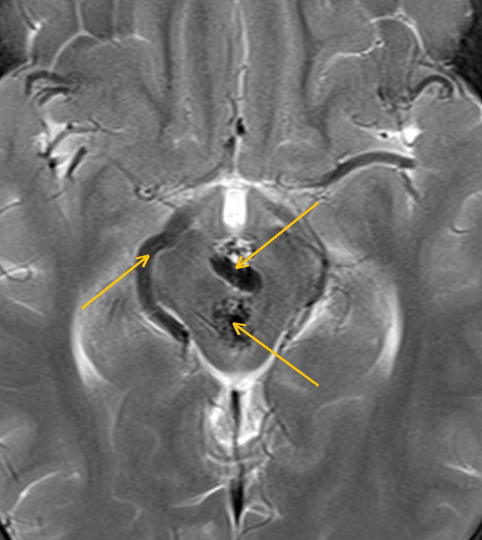
The striking finding is on T2. Deep in the middle of the brain, where the third ventricle, the hypothalamus and the top of the midbrain meet, there is a cluster of black, worm-like shapes. These are flow voids: fast-moving blood gives no signal, so abnormal vessels look like dark tubes and dots.
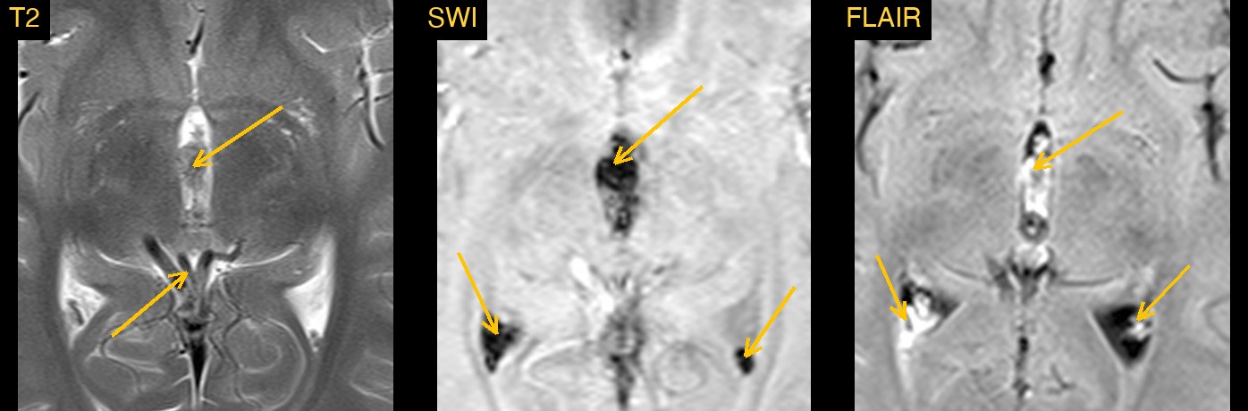
The second finding is blood. The third ventricle is filled with material that does not look like normal fluid, and blood products lie at the back of the lateral ventricles.
How Monitraq's AI read the scan
- It described the vessels: a cluster of serpiginous and punctate T2 flow voids filling the third ventricle and hypothalamic region and extending into the interpeduncular and perimesencephalic cisterns, about 10 x 20 mm. It noted prominent round flow voids along the right side of the midbrain, tortuous dark vessels near the vein of Galen, and serpiginous enhancing vessels after contrast.
- It worked out the blood: the material in the third ventricle was intermediate on T2, bright on FLAIR, partly bright on the unenhanced T1 and blooming on SWI, which it read as subacute clot. Layering FLAIR-bright, SWI-dark material in the occipital horns and atria meant intraventricular haemorrhage.
- It checked for pressure: the lateral ventricles were not enlarged, the temporal horns were only mildly visible and there was no midline shift.
- It gave a ranked differential: a deep (diencephalic or choroidal) arteriovenous malformation with intraventricular haemorrhage as the leading diagnosis; a haemorrhagic cavernous malformation, or a haemorrhagic third ventricular or suprasellar tumour (germ cell tumour, choroid plexus tumour, craniopharyngioma), as possible alternatives; and a dural arteriovenous fistula as unlikely.
- It flagged the scan as urgent and listed next steps: urgent neurosurgical and neurovascular referral; CT or MR angiography followed by catheter angiography to map the feeding arteries, draining veins and any aneurysm; watching for hydrocephalus, with a CT head if conscious level falls or headache worsens; and, if angiography showed no shunt, a contrast MRI once the blood had cleared.
It also stated what the study could not show. Without angiographic images, the feeding arteries and draining veins could not be mapped on MRI alone.
Dr Priti, the radiologist who reviewed this case, reported a midbrain arteriovenous malformation with serpiginous flow voids and enlarged feeding vessels in the interpeduncular and quadrigeminal region, with a secondary intraventricular haemorrhage.
What is a brain arteriovenous malformation?
A brain AVM is a tangle of abnormal vessels, called the nidus, where arteries drain straight into veins without a normal capillary bed in between. Blood flows through it fast and under high pressure.
Bleeding is the most common way an AVM comes to light. Others present with seizures, headache or a focal deficit, and some are found by chance. Deep AVMs, near the thalamus, basal ganglia, third ventricle or brainstem, often bleed into the ventricles. Deep location and deep venous drainage are both recognised as features that make treatment harder.
Key MRI signs of a brain AVM
- A cluster of serpiginous flow voids on T2, often called a "bag of worms".
- Enlarged feeding arteries and dilated draining veins leading to and from the cluster.
- Little or no mass effect from the nidus itself.
- Blood products of different ages, best seen as dark blooming on SWI.
- Serpiginous enhancing vessels after contrast.
- Bleeding into the brain, the ventricles or the subarachnoid space.
Differential diagnosis
- Cavernous malformation (cavernoma). A "popcorn" lesion with a complete dark haemosiderin rim. It has no enlarged feeding vessels or flow voids and does not show on angiography.
- Haemorrhagic tumour. A solid enhancing part separate from the vessels points to a tumour. Repeat imaging after the blood clears can help.
- Dural arteriovenous fistula. The shunt lies in the dura, with no nidus in the brain itself. Angiography shows dural arterial feeders.
Frequently asked questions
What does an AVM look like on MRI?
On T2 it shows as a tangle of dark flow voids, with enlarged vessels running to and from it. After contrast the vessels enhance, and SWI shows any old or recent bleeding as dark areas.
Can MRI alone confirm an AVM?
MRI can strongly suggest it, but catheter angiography is the reference test. It maps the feeding arteries, the nidus, the draining veins and any aneurysm.
Why does blood show up on SWI and FLAIR?
Blood products disturb the local magnetic field, so SWI shows them as dark blooming areas. On FLAIR, blood in the ventricles is brighter than the normally dark CSF.
A draft for the radiologist
Here the AI put the bleeding and its suspected vascular cause first, marked the study urgent and set out the angiography needed to confirm it.
It gives a draft, not a diagnosis. The AI flags, describes and suggests. A radiologist confirms, corrects and signs.
Monitraq's AI produces draft reports to support qualified radiologists. It is not a diagnosis and is not intended for use by patients. All case details have been removed or generalised to protect patient privacy, and the images have been cropped and stripped of identifying information.