Cholesteatoma on MRI with non-EPI DWI: how Monitraq's AI read it
A cholesteatoma of the middle ear can be shown on MRI with one sequence above all others: non-EPI diffusion-weighted imaging. In this brain MRI a single bright spot in the right middle ear and mastoid made the case. Here is how Monitraq's AI worked through it.
MRI findings
The study was a brain MRI with coronal FLAIR and axial FLAIR after contrast, SWI, axial echo-planar DWI with an ADC map, a coronal non-echo-planar (non-EPI) DWI, a thin 3D T2 (CISS) and fat-saturated T1 after contrast. No clinical question was given.
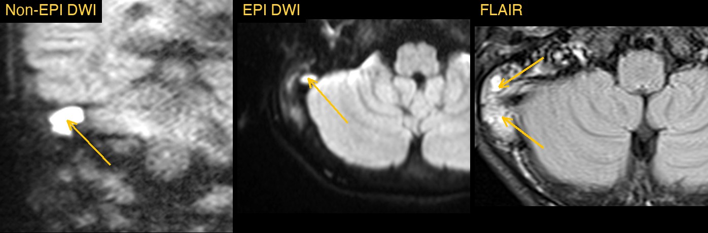
The brain itself looks normal. The finding is in the right temporal bone. The air cells of the right mastoid and the right middle ear are filled with soft tissue that is bright on FLAIR, while the left mastoid is still full of air, which shows as black.
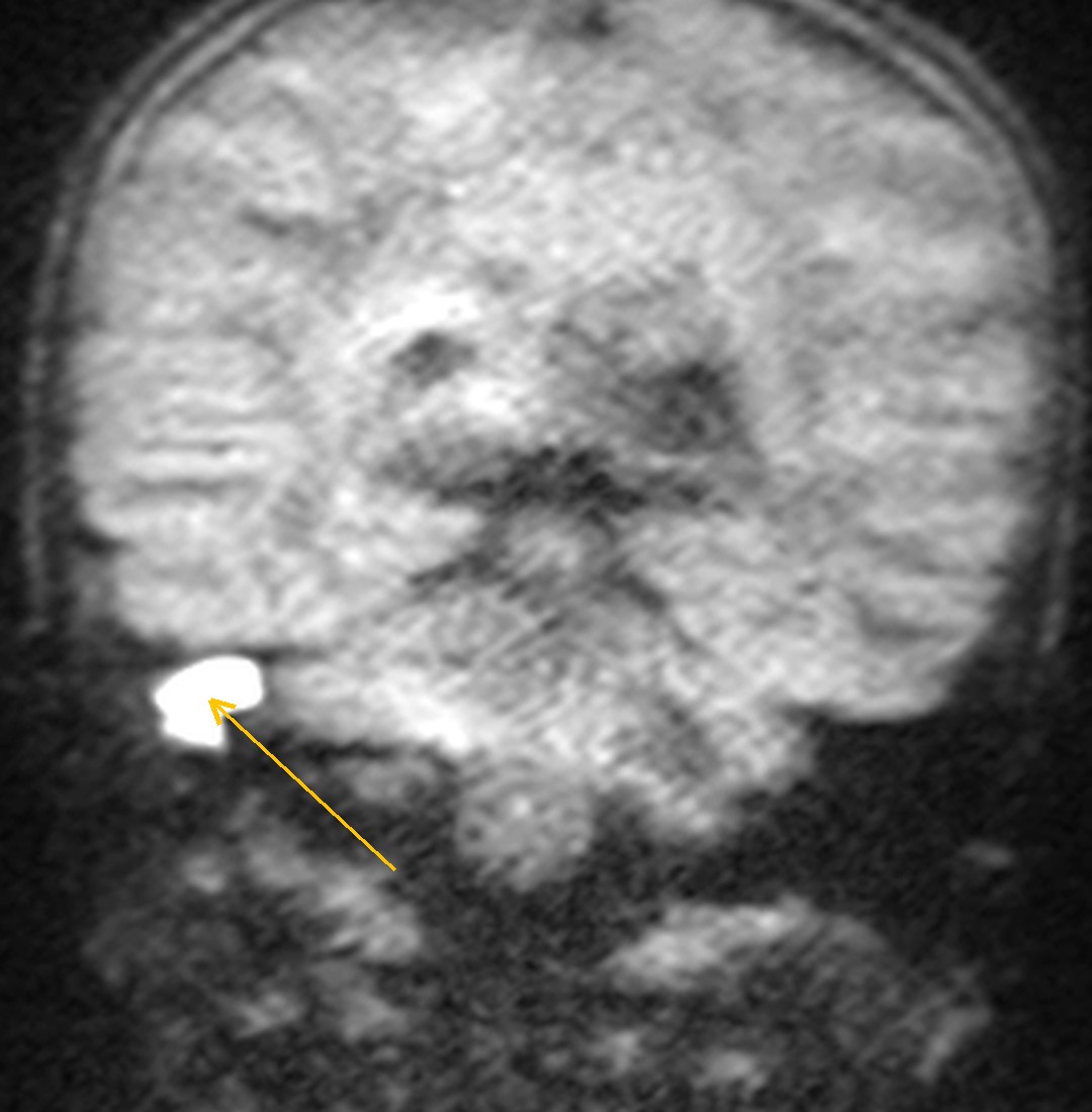
Within that soft tissue sits a well-defined focus that is very bright on non-EPI DWI and stays bright over several neighbouring coronal slices. It is also bright on the standard echo-planar DWI.
How Monitraq's AI read the scan
- It found the side and the space: soft tissue filling the right mastoid air cells and middle ear, bright on FLAIR, with a normally aerated left mastoid.
- It singled out the key sign: a well-defined focus, markedly bright on non-EPI (HASTE) DWI over several contiguous coronal slices and bright on axial EPI DWI, measuring about 13 x 9 mm.
- It checked for spread inside the skull: the adjacent temporal lobe, cerebellum and dura were normal, with no collection, abscess or venous sinus thrombosis.
- It gave a ranked differential: cholesteatoma (primary, or residual or recurrent after surgery) as the leading diagnosis; otomastoiditis with fluid and granulation tissue as possible; and cholesterol granuloma and glomus tympanicum as unlikely.
- It set out next steps: high-resolution CT of the temporal bones to assess erosion of the ossicles, scutum, tegmen and labyrinth; ENT review with otoscopy and a history of any previous ear surgery; and, if CT and the clinical picture were equivocal, a repeat non-EPI DWI with an ADC map.
It also stated what the study could not show. The AI reported that the cochlea, vestibule and semicircular canals were not clearly shown on the images it reviewed, so it did not assess the inner ear. Its CT recommendation included the labyrinth.
Dr Priti, the radiologist who reviewed this case, reported a cholesteatoma of the right middle ear, bright on non-EPI DWI, with inner ear involvement: loss of the normal fluid signal in the right cochlea and semicircular canals (labyrinthitis).
What is a cholesteatoma?
A cholesteatoma is a growth of keratinising squamous epithelium in the middle ear or mastoid. It is not a true tumour, but it slowly expands and erodes bone. Most are acquired, often linked to a retraction pocket of the eardrum. Some are present from birth, and some come back after surgery.
Typical symptoms are a smelly, persistent ear discharge and hearing loss. If it keeps growing, it can erode the ossicles, the bony covering of the inner ear, the facial nerve canal and the roof of the middle ear. Rarely it spreads into the skull.
CT shows the bone erosion. MRI shows what the soft tissue is. The keratin inside a cholesteatoma restricts the movement of water, so it is bright on diffusion imaging, while fluid and inflamed tissue usually are not.
Key MRI signs of cholesteatoma
- Marked brightness on non-EPI DWI, the most specific MRI sign.
- Low signal on the ADC map, confirming restricted diffusion.
- No central enhancement after contrast. Only the rim or nearby inflamed tissue enhances.
- Intermediate to low T1 and moderately bright T2 signal.
- Nearby fluid or inflamed mucosa in the mastoid, which does not restrict diffusion.
Differential diagnosis
- Otomastoiditis with granulation tissue. The content behaves like fluid, very bright on T2 with a high ADC, and the granulation tissue enhances. There is no bright non-EPI DWI focus.
- Cholesterol granuloma. Bright on both plain T1 and T2, without restricted diffusion.
- Glomus tympanicum. A small, avidly enhancing nodule on the cochlear promontory, with no restricted diffusion.
- Abscess. Also restricts diffusion, but has an enhancing wall and usually a clear clinical picture of infection.
Frequently asked questions
Which MRI sequence is best for cholesteatoma?
Non-echo-planar DWI. It has fewer artefacts near the air and bone of the temporal bone than standard echo-planar DWI, so small cholesteatomas show more clearly.
What does cholesteatoma look like on DWI?
It is a well-defined bright focus on DWI, with low signal on the ADC map. Fluid and granulation tissue around it are usually not bright on DWI.
Why is CT still needed if MRI shows a cholesteatoma?
High-resolution CT shows the bone. It is the test for erosion of the ossicles, the scutum, the tegmen and the bony labyrinth, which guides surgery.
A draft for the radiologist
Here the AI found the one sign that mattered most, put cholesteatoma first and named what CT and ENT review still had to answer. The radiologist's report also describes the inner ear, the kind of detail a radiologist settles before signing.
It gives a draft, not a diagnosis. The AI flags, describes and suggests. A radiologist confirms, corrects and signs.
Monitraq's AI produces draft reports to support qualified radiologists. It is not a diagnosis and is not intended for use by patients. All case details have been removed or generalised to protect patient privacy, and the images have been cropped and stripped of identifying information.