Fibrous dysplasia of the sphenoid wing on MRI: how Monitraq's AI read it
Fibrous dysplasia of the sphenoid wing can look unusual on MRI, because MRI is not the test that shows it best. This scan had only T2 images, yet the expanded, bubbly bone beside the orbit was clear. Here is how Monitraq's AI read it.
MRI findings
A patient had a brain MRI with fat-saturated T2 images in two planes, axial and coronal, plus a low-resolution localiser. There were no T1, FLAIR, diffusion or contrast images.
The right greater wing of the sphenoid bone, the part that forms the outer wall of the orbit and the front of the middle cranial fossa, was expanded and wider than the left. Instead of the normal dark bone, it was filled with an intermediate T2 matrix holding many small, well-defined bright pockets, giving a bubbly or honeycomb look. One larger rounded pocket bulged towards the back of the orbit. The lesion touched the right temporal pole and the outer wall of the right orbit, but there was no swelling in the brain and no separate soft-tissue mass.
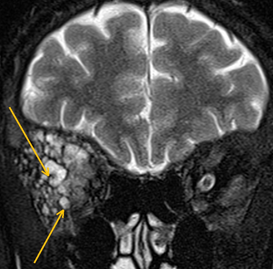
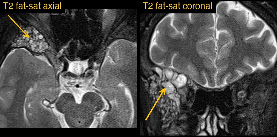
How Monitraq's AI read the scan
- It placed the lesion in bone: a single expansile lesion replacing and widening the right greater sphenoid wing and lateral orbital wall, reaching the front of the right middle cranial fossa and the temporal fossa.
- It described the texture: a heterogeneous intermediate T2 matrix with numerous small, well-defined bright locules, a bubbly or honeycomb pattern, with one larger locule bulging towards the orbital apex.
- It checked the neighbours: the lesion abutted the right temporal pole and the orbit without brain oedema or a separate soft-tissue mass. The right globe appeared slightly forward of the left, possible mild proptosis, though head rotation contributed.
- It gave a ranked differential: fibrous dysplasia was favoured; intraosseous haemangioma or venous malformation and aneurysmal bone cyst were the main alternatives.
- It said what the study could not show, with no T1, diffusion or contrast images, and listed next steps: thin-section CT of the skull base and orbits to look for a ground-glass matrix, cortical integrity and narrowing of the orbit and nerve canals; contrast-enhanced MRI if CT is not diagnostic; and an eye assessment for proptosis, vision and optic nerve function.
It also reported a few small, chronic white matter spots and two incidental findings, a partly empty sella and a small cyst in the scalp, and kept them in the background.
Dr Priti, the radiologist who reviewed this case, reported fibrous dysplasia as the differential diagnosis.
What is fibrous dysplasia?
Fibrous dysplasia is a benign developmental bone disorder. Normal bone is replaced by a mix of fibrous tissue and immature, poorly formed bone. It can affect one bone (monostotic) or several (polyostotic). When several bones are involved together with skin pigmentation and hormone problems, it is part of McCune-Albright syndrome.
The skull and facial bones are common sites. There the expanding bone can change the shape of the face or skull, push the eye forward, or narrow the canals that carry nerves, including the optic nerve. Many lesions cause no symptoms and are found by chance.
Key MRI signs of fibrous dysplasia
- An expanded bone with its outer shape preserved, rather than a destructive mass.
- Variable signal: often low to intermediate on T1 and mixed on T2, sometimes with cystic, fluid-bright areas.
- Variable enhancement after contrast, which can make it look like a tumour.
- No brain oedema and no soft-tissue mass breaking out of the bone.
- CT is the key test: a ground-glass matrix within expanded bone is the classic sign.
Differential diagnosis
On MRI alone, fibrous dysplasia can resemble other bone lesions. The AI's alternatives were intraosseous haemangioma or venous malformation, which can also look bright and lobulated, and aneurysmal bone cyst, which tends to show fluid-filled spaces, sometimes with fluid levels. More generally, other sphenoid wing lesions include meningioma with bone thickening and metastasis. CT usually settles the question, which is why the AI made it the first recommendation.
Frequently asked questions
Why is CT better than MRI for fibrous dysplasia?
CT shows the internal bone pattern directly. The typical ground-glass matrix within expanded bone is seen far better on CT than on MRI, where signal is variable.
What does fibrous dysplasia look like on T2 MRI?
It is variable. It is often intermediate to low, but areas of cystic change can be bright, and the overall look can be mixed or bubbly, as in this case.
Can fibrous dysplasia of the sphenoid wing affect vision?
Yes. Expanding bone around the orbit or the optic canal can push the eye forward or narrow the canal around the optic nerve, so eye assessment is often advised.
A draft for the radiologist
A limited study still deserves a careful read. Here the AI described the lesion precisely, said plainly which sequences the study did not include, and pointed to the test most likely to give the answer.
It gives a draft, not a diagnosis. The AI flags, describes and suggests. A radiologist confirms, corrects and signs.
Monitraq's AI produces draft reports to support qualified radiologists. It is not a diagnosis and is not intended for use by patients. All case details have been removed or generalised to protect patient privacy, and the images have been cropped and stripped of identifying information.