Focal cortical dysplasia on MRI: how Monitraq's AI read a left frontal lesion
Focal cortical dysplasia on MRI is often subtle, and it is one of the most important findings to get right in epilepsy imaging. This scan showed a small bright patch in the left frontal lobe. Here is how Monitraq's AI described it, and what separates it from a tumour.
MRI findings
A patient had a brain MRI with T2, FLAIR, diffusion, susceptibility and T1 images. No clinical history came with the scan.
In the left frontal lobe, in the superior and middle frontal gyri, there was a single ill-defined lesion involving the cortex and the white matter just beneath it. It was bright on T2 and FLAIR. The cortex there looked thickened, and the normally sharp line between grey and white matter was blurred. The lesion did not push on anything: the sulci above it and the frontal horn of the ventricle nearby were preserved, and there was no swelling around it.
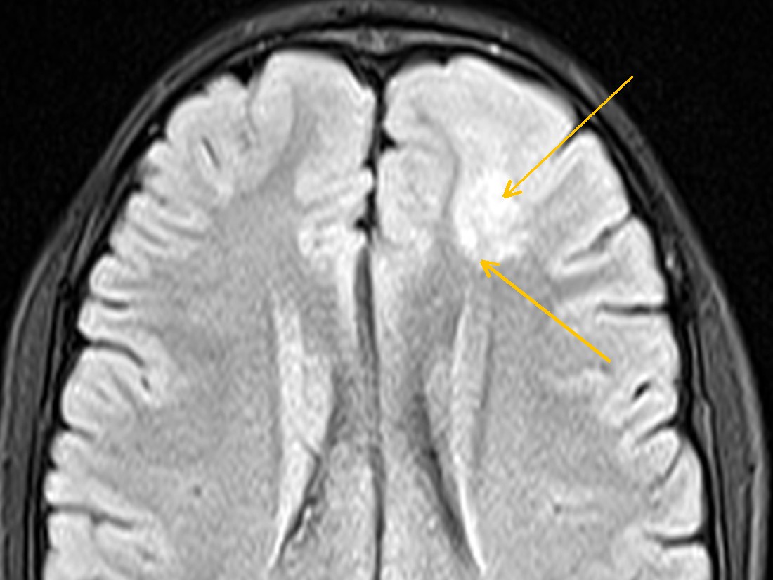
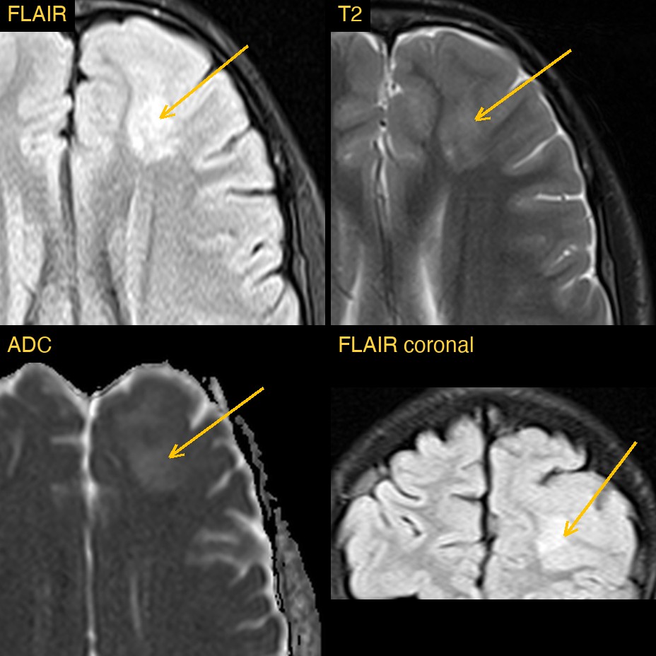
How Monitraq's AI read the scan
- It placed the lesion: a single cortical-subcortical lesion in the left superior and middle frontal gyri, about 20 by 25 mm on axial FLAIR.
- It named the dysplasia signs: T2/FLAIR brightness with cortical thickening and blurring of the grey-white junction.
- It listed what was absent: no restricted diffusion (the lesion was mildly bright on ADC), no bubbly or multicystic component, no blooming on SWI, no definite enhancement, no oedema and no mass effect.
- It gave a ranked differential: the appearance was suggestive of focal cortical dysplasia (type II). A low-grade glial or glioneuronal tumour (low-grade glioma, ganglioglioma or DNET) was the main alternative.
- It listed next steps: clinical and EEG correlation for focal seizures from the left frontal lobe; a dedicated epilepsy-protocol MRI with thin-section 3D FLAIR and coronal T2 through the frontal lobes and hippocampi, including a search for a transmantle band; MR spectroscopy to help separate dysplasia from tumour; and a follow-up MRI to confirm stability.
Dr Priti, the radiologist who reviewed this case, reported focal cortical dysplasia type IIb: a left frontal cortical-subcortical lesion with a transmantle sign, without the typical bubbly appearance of a DNET.
What is focal cortical dysplasia?
Focal cortical dysplasia (FCD) is a malformation of cortical development. During development, a small area of cortex forms abnormally: its layers are disorganised and it may contain abnormal nerve cells. It is one of the most common causes of focal epilepsy that does not respond to medication, in both children and adults.
FCD is grouped into types. In type II, the cortex contains large abnormal neurons called dysmorphic neurons. Type IIb also contains balloon cells, and it is the type most often visible on MRI. Finding it matters because a well-defined lesion that matches the seizure onset may be suitable for epilepsy surgery.
Key MRI signs of focal cortical dysplasia
- Focal cortical thickening.
- Blurring of the grey-white matter junction.
- Bright T2/FLAIR signal in the subcortical white matter beneath the abnormal cortex.
- The transmantle sign: a thin, funnel-shaped band of FLAIR brightness running from the cortex towards the lateral ventricle, typical of type IIb.
- Usually no mass effect, no oedema, no restricted diffusion and no enhancement.
- Thin-section 3D FLAIR and T1 images improve detection of small lesions.
Differential diagnosis
The main question is dysplasia versus a low-grade tumour. A dysembryoplastic neuroepithelial tumour (DNET) is also cortical and associated with epilepsy, but it usually has a bubbly, multicystic look, sometimes with a bright rim on FLAIR. Ganglioglioma often has a cyst and an enhancing nodule, and may contain calcium. A low-grade glioma tends to expand the gyrus and cause some mass effect. In this case the AI noted no bubbly component, no enhancement and no mass effect, but it still kept a low-grade tumour as the main alternative.
Frequently asked questions
What is the transmantle sign in focal cortical dysplasia?
It is a thin band of T2/FLAIR brightness that tapers from the abnormal cortex down towards the lateral ventricle. It reflects the path of abnormal cells and is a typical sign of FCD type IIb.
Which MRI sequence shows focal cortical dysplasia best?
FLAIR, ideally as thin-section 3D FLAIR, is the most sensitive for the subcortical signal and the transmantle sign. T1-weighted 3D images help to show cortical thickening and grey-white blurring.
How do you tell focal cortical dysplasia from a DNET on MRI?
A DNET usually looks bubbly or multicystic and may have a bright rim on FLAIR. FCD typically shows cortical thickening, grey-white blurring and sometimes a transmantle band, without a cystic component.
A draft for the radiologist
In epilepsy imaging the finding can be small and quiet, and the report has to say what is there, what is not, and which scan will show it better. Monitraq's AI is built to set out that reasoning so the radiologist can check it quickly.
It gives a draft, not a diagnosis. The AI flags, describes and suggests. A radiologist confirms, corrects and signs.
Monitraq's AI produces draft reports to support qualified radiologists. It is not a diagnosis and is not intended for use by patients. All case details have been removed or generalised to protect patient privacy, and the images have been cropped and stripped of identifying information.