Large ventricles in an infant: how Monitraq's AI reads a pattern that points to congenital CMV
An infant had a brain MRI. The ventricles were large, the white matter around them was abnormal and the corpus callosum was thin. Each finding has several possible causes on its own. Together they point to one. Here is how Monitraq's AI worked through the scan.
The scan
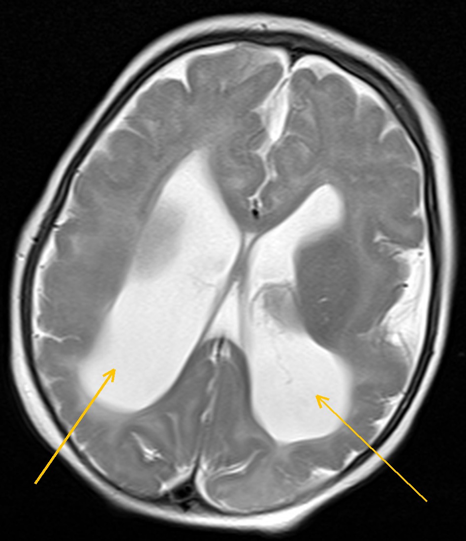
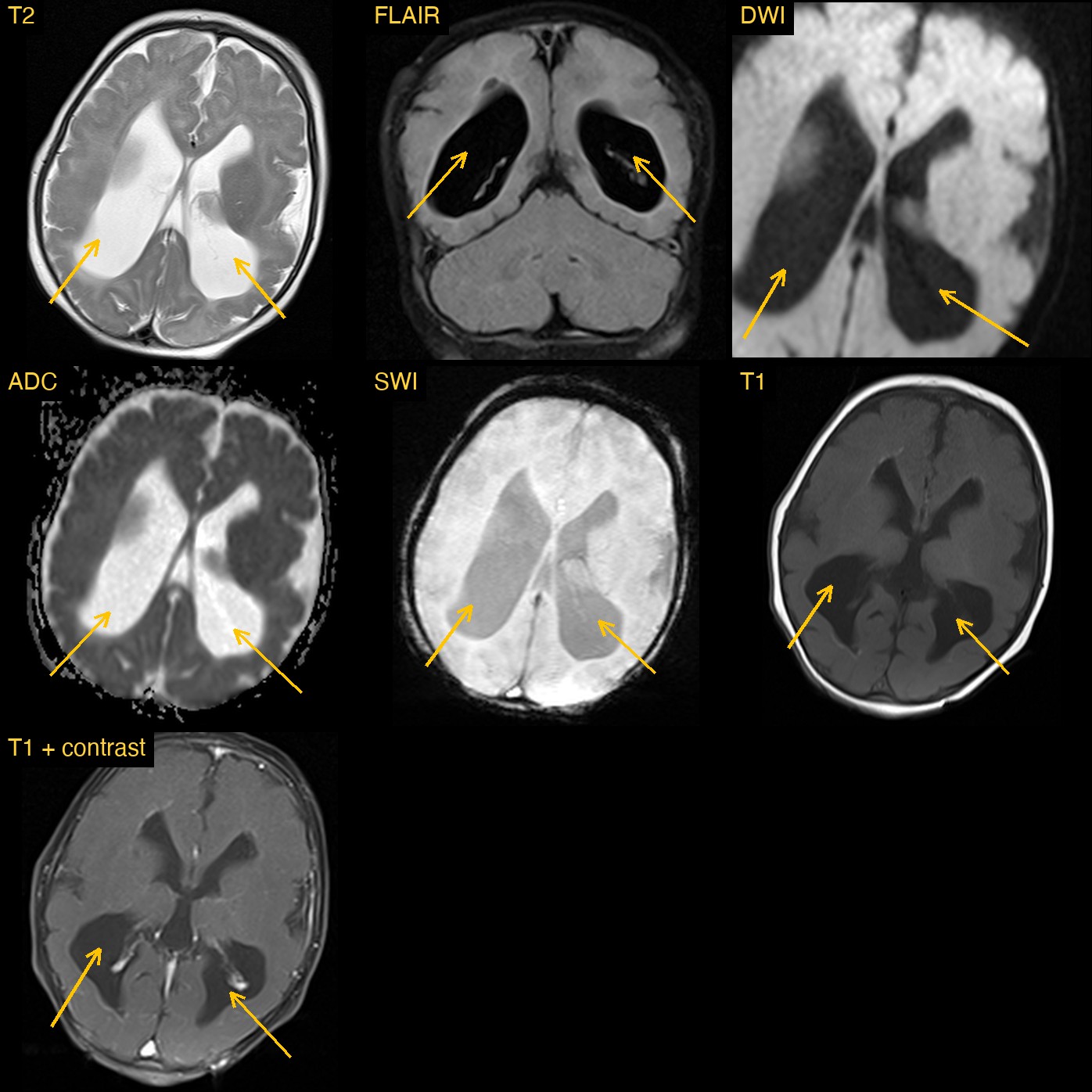
The most obvious finding was the size of the lateral ventricles, the fluid spaces in the middle of the brain. They were enlarged, and not evenly: the back parts (the atria and occipital horns) were much larger than the front. This shape is called colpocephaly. The temporal horns were dilated too.
The white matter told the rest of the story. On T2 and FLAIR images there was hazy brightness around the ventricles, and there was less white matter than expected around their back parts. The corpus callosum, the large band of fibres that joins the two halves of the brain, was thin and stretched.
Why the pattern matters more than each finding
Large ventricles alone can come from blocked fluid flow, from brain tissue that never developed fully, or from tissue that was lost. White matter change around the ventricles has its own list of causes. A report that lists each finding separately is accurate, but it leaves the most useful step undone: asking what single process could explain them all.
How Monitraq's AI read the scan
- It described the ventricles precisely: enlarged lateral ventricles with a colpocephalic shape, disproportionately large atria and occipital horns, and dilated temporal horns. The fourth ventricle was normal.
- It described the white matter: hazy periventricular T2 and FLAIR brightness around the atria and bodies of the ventricles, reduced white matter around the atria, and a thin, stretched corpus callosum.
- It put the findings together: ventriculomegaly, periventricular white matter change, white matter volume loss and a thin corpus callosum, taken as one pattern, raised congenital cytomegalovirus (CMV) or another TORCH infection as the leading consideration.
- It weighed the alternatives: chronic periventricular white matter injury was named as the main alternative, and it advised that hydrocephalus from blocked fluid flow be excluded clinically.
- It listed the next steps: congenital CMV testing on the newborn dried blood spot and a TORCH screen; hearing and eye assessment; a review of the full susceptibility images and consideration of low-dose CT to look for calcification around the ventricles; and a check of the head circumference trend and serial imaging to judge whether the ventricles are growing.
Dr Priti, the radiologist who reviewed this case, reported congenital CMV infection: enlarged ventricles with irregular margins, periventricular white matter changes and a thin corpus callosum, all brought together into one diagnosis.
What is congenital CMV infection?
Cytomegalovirus is a common virus that usually causes mild or no illness in healthy adults. If a mother is infected during pregnancy, the virus can cross the placenta and infect the baby. Congenital CMV is the most common congenital viral infection and a leading non-genetic cause of hearing loss in children.
The virus can damage the developing brain. On imaging this can show as enlarged ventricles, white matter abnormality, calcifications around the ventricles, cysts and malformations of the cortex. Many babies have no signs at birth, so imaging findings can be an important clue. Confirming the infection needs laboratory testing, and early recognition matters because affected children need hearing and developmental follow-up.
A draft for the radiologist
A good radiologist does not stop at listing findings. They ask which single diagnosis explains them all. Monitraq's AI is built to do the same, and to show its reasoning so the radiologist can check it quickly.
It gives a draft, not a diagnosis. The AI flags, describes and suggests. A radiologist confirms, corrects and signs.
Monitraq's AI produces draft reports to support qualified radiologists. It is not a diagnosis and is not intended for use by patients. All case details have been removed or generalised to protect patient privacy, and the image has been cropped and stripped of identifying information.