A worn disc at the bottom of the spine: how Monitraq's AI reads L5-S1
A lumbar spine MRI showed the lowest disc, between the last lumbar vertebra and the sacrum, worn down and dark, with a small rounded piece pushing backwards towards the nerves. Here is how Monitraq's AI worked through the scan.
The scan
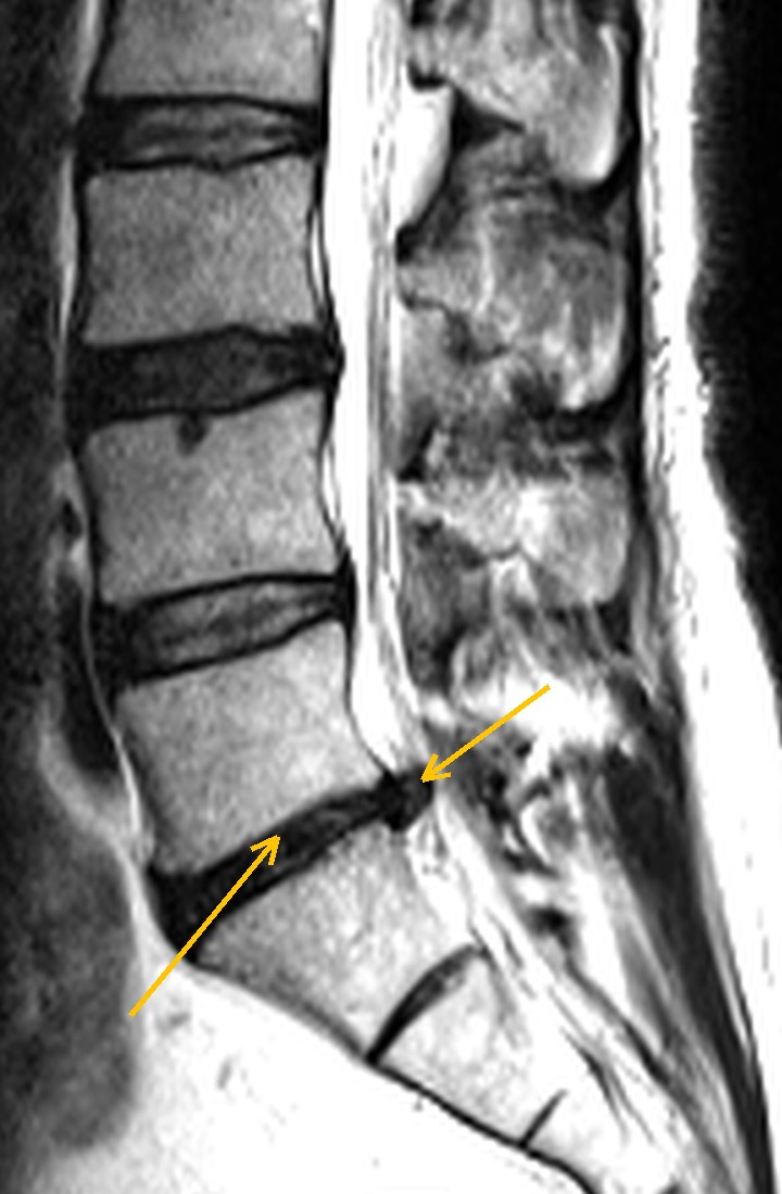
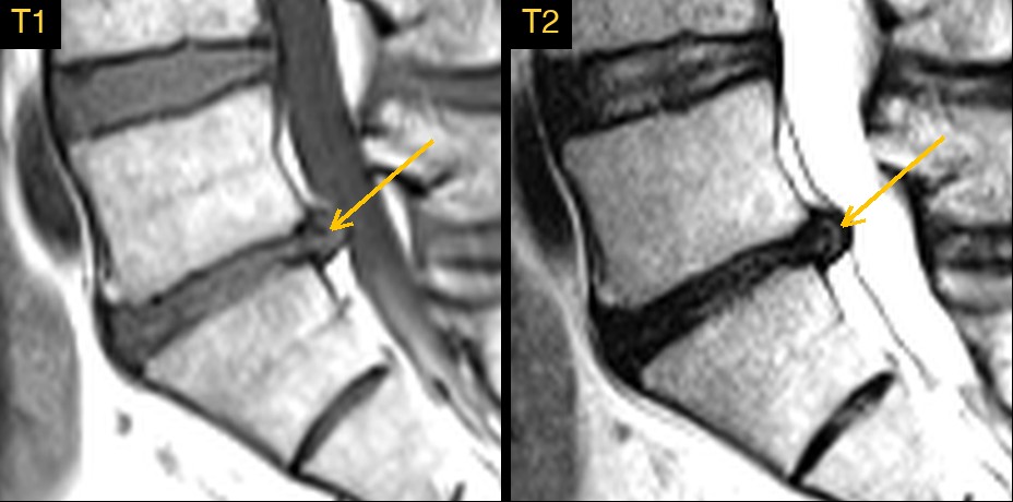
This lumbar spine MRI comes from a public research collection. It had sagittal T1 and T2 images, which show the spine from the side. On T2, a healthy disc has a bright, water-rich centre; a worn disc loses that water and turns dark.
The disc at L5-S1, the lowest level, was dark from front to back and thinner than it should be. At its back edge, a small rounded bump stood out and pressed on the front of the fluid sac that holds the nerve roots.
Why the detail matters
Many adults have some disc wear on MRI, and much of it causes no symptoms. What helps the treating doctor is a clear answer to a few questions: which level, how worn, whether disc material is pushing out focally, which way it points, and whether it is close to a nerve root. A vague "degenerative changes" does not answer them.
How Monitraq's AI read the scan
- It graded the worst disc: L5-S1 was uniformly dark on T2, with complete loss of its normal bright centre and clear loss of height, most marked at the back.
- It found the focal protrusion: a focal, rounded, T2-dark protrusion that stood out from the rest of the L5-S1 disc margin and indented the front of the thecal sac. It was seen on the midline and slightly right-of-midline slices.
- It described its direction and effect: small, central to slightly right paracentral, with fluid (CSF) still preserved behind it and no significant narrowing of the central canal.
- It was clear about what the images could not show: without axial images it could not confirm whether the protrusion touched the descending S1 nerve roots, or exactly which way it pointed.
- It listed the next steps: obtain or review axial T2 images through L4 to S1 to assess the protrusion, the S1 roots and the lateral recesses; correlate clinically for S1 nerve root symptoms; and confirm the level numbering against a whole-spine localiser if any intervention is planned.
The expert gradings that come with this public dataset record a disc herniation at L5-S1 and degenerative disc disease, worst at Pfirrmann grade 5.
What is a disc protrusion?
Each intervertebral disc has a soft, water-rich centre (the nucleus pulposus) held in by a tough outer ring (the annulus fibrosus). With age and wear the nucleus dries out and darkens on T2, and the disc loses height. Radiologists often grade this on the Pfirrmann scale from 1 to 5, where grade 5 is a dark, collapsed disc.
A herniation is disc material pushed beyond the normal edge of the disc over a limited part of its circumference. When the base of the displaced material is wider than its tip, it is called a protrusion. At L5-S1, a central or paracentral herniation usually affects the S1 nerve root, which can cause pain or numbness down the back of the leg into the sole of the foot and a reduced ankle reflex.
A draft for the radiologist
A good radiologist names the level, describes the disc and the protrusion precisely, and says plainly what the images cannot settle. Monitraq's AI is built to do the same, and to show its reasoning so the radiologist can check it quickly.
It gives a draft, not a diagnosis. The AI flags, describes and suggests. A radiologist confirms, corrects and signs.
Monitraq's AI produces draft reports to support qualified radiologists. It is not a diagnosis and is not intended for use by patients. All case details have been removed or generalised to protect patient privacy, and the image has been cropped and stripped of identifying information. Image: SPIDER lumbar spine MRI dataset, CC BY 4.0.