Bright spots and a cerebellar lesion: how Monitraq's AI reads a scan that suggests NF1
A brain MRI arrived with no clinical history. It showed small bright spots deep in the brain and a rounded lesion in the cerebellum. Read together, the images pointed to a genetic condition, neurofibromatosis type 1. Here is how Monitraq's AI worked through the scan.
The scan
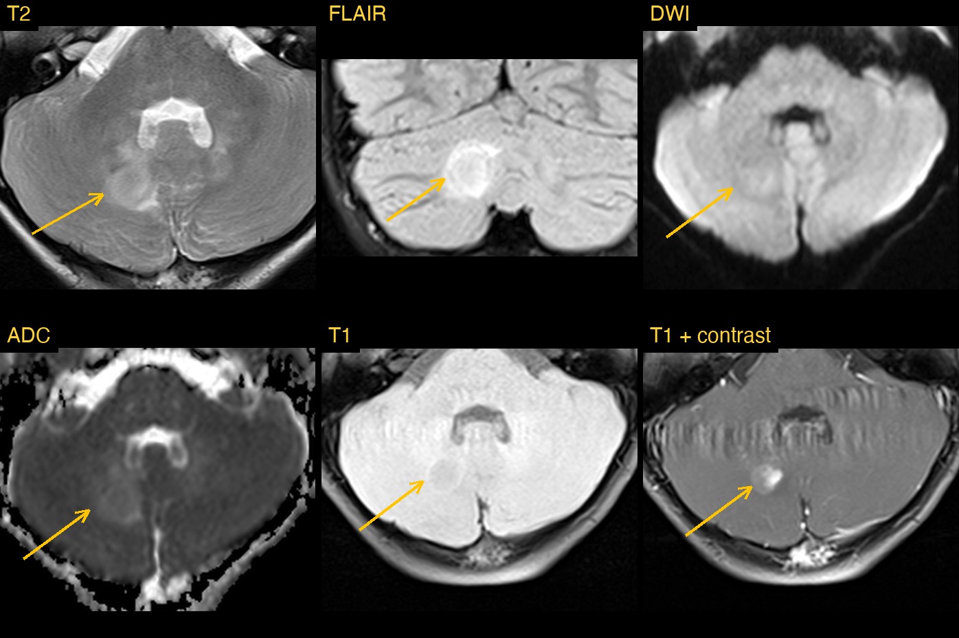
On T2 and FLAIR images there were rounded bright foci in both globi pallidi, part of the deep grey matter, larger on the left. They caused no mass effect and did not restrict diffusion. The deep cerebellar nuclei, the dentate nuclei, were bright on both sides as well.
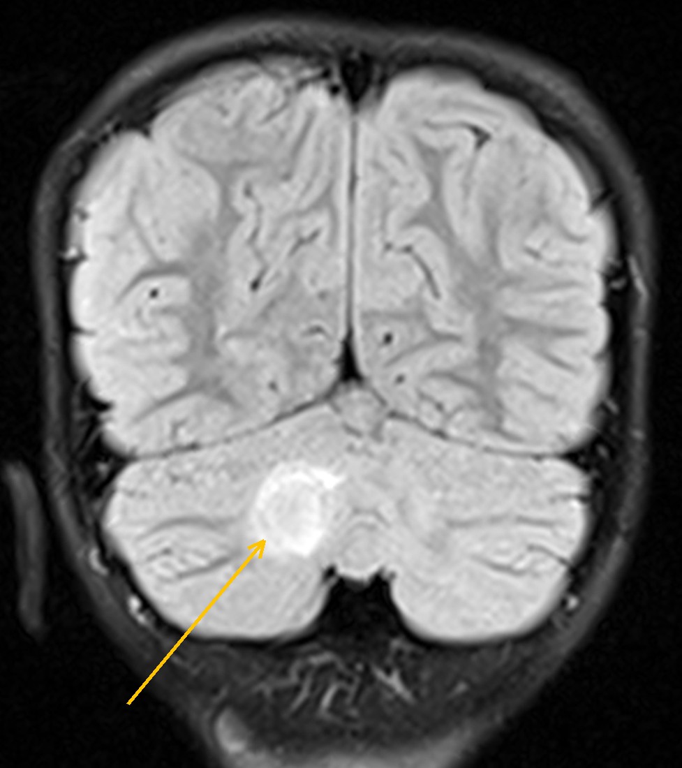
In the right cerebellar hemisphere, next to the midline, the change was bigger: a rounded lesion of about 15 mm with a target-like rim on coronal FLAIR and a little surrounding signal. It did not press on the fourth ventricle and there was no hydrocephalus.
Why the whole scan matters
A single bright lesion in the cerebellum has a long list of possible causes. The small bright spots in the globi pallidi and dentate nuclei are easy to treat as a separate, minor finding. But that combination is a recognised imaging signature of neurofibromatosis type 1, and once it is recognised, the cerebellar lesion is read in a different light.
How Monitraq's AI read the scan
- It described the deep grey matter foci: rounded T2/FLAIR-bright foci in both globi pallidi, left larger than right, with mild T1 brightness, no mass effect, no restricted diffusion and no enhancement on the sampled slices.
- It looked at the cerebellum carefully: both dentate regions were brighter than cortex on T2 and FLAIR, without restricted diffusion. On the right the change formed a larger, rounded, rim-like lesion of about 15 mm, with mild surrounding oedema and a small enhancing part.
- It linked the findings: the pallidal and dentate foci were typical of NF1 focal areas of signal intensity (FASI), even though no history of NF1 had been given.
- It weighed the cerebellar lesion in that light: because it was larger, rounded and partly enhancing, it could be an atypical FASI or a low-grade glioma such as a pilocytic astrocytoma. The AI favoured a low-grade glioma and said that post-contrast and serial imaging are needed to tell them apart.
- It listed the next steps: clinical assessment for NF1 (skin, eyes including Lisch nodules and vision, family history) and consideration of genetic testing; a short-interval follow-up MRI with contrast to see whether the cerebellar lesion grows; and dedicated imaging of the optic pathways if NF1 is confirmed.
Dr Priti, the radiologist who reviewed this case, reported neurofibromatosis type 1: focal areas of signal intensity (FASI) in the globi pallidi, dentate nuclei and cerebellum, with a cerebellar lesion that could be an atypical FASI or a low-grade glioma.
What are FASI in NF1?
Neurofibromatosis type 1 is a common inherited condition. It is known for skin signs such as café-au-lait spots and neurofibromas, and it also affects the brain and eyes. Many children with NF1 have small bright spots on T2 and FLAIR MRI called focal areas of signal intensity (FASI), also known as unidentified bright objects. They are typically found in the globi pallidi, cerebellum, brainstem and thalami, cause no mass effect, do not enhance, and often fade with age.
People with NF1 also have a higher risk of brain tumours, mostly low-grade gliomas such as optic pathway gliomas and pilocytic astrocytomas. A spot that is larger than usual, enhances or presses on its surroundings is therefore watched closely with follow-up imaging.
A draft for the radiologist
A good radiologist reads every finding in the context of the others, especially when no history comes with the scan. Monitraq's AI is built to do the same, and to show its reasoning so the radiologist can check it quickly.
It gives a draft, not a diagnosis. The AI flags, describes and suggests. A radiologist confirms, corrects and signs.
Monitraq's AI produces draft reports to support qualified radiologists. It is not a diagnosis and is not intended for use by patients. All case details have been removed or generalised to protect patient privacy, and the image has been cropped and stripped of identifying information.