Small MCA branch stroke on MRI: how Monitraq's AI read it
A small MCA branch stroke on MRI can be easy to pass over in a brain that also shows generalised volume loss. This adult's scan held an old infarct behind the left Sylvian fissure. Here is how Monitraq's AI found it, described it and set out what the images could not show.
MRI findings
This brain MRI comes from a public research collection. Only one kind of image was available: a T2-weighted volume, viewed in the axial, coronal and sagittal planes. On T2, water and fluid look bright.
Behind the left Sylvian fissure, in the parietal lobe, there was a patch of brighter tissue. It involved the cortex and the white matter beneath it and reached towards the side of the left lateral ventricle. Nothing was pushed aside. Instead, the nearby fissure and the back of the left ventricle were wider than expected, as if the brain had shrunk back from that spot.
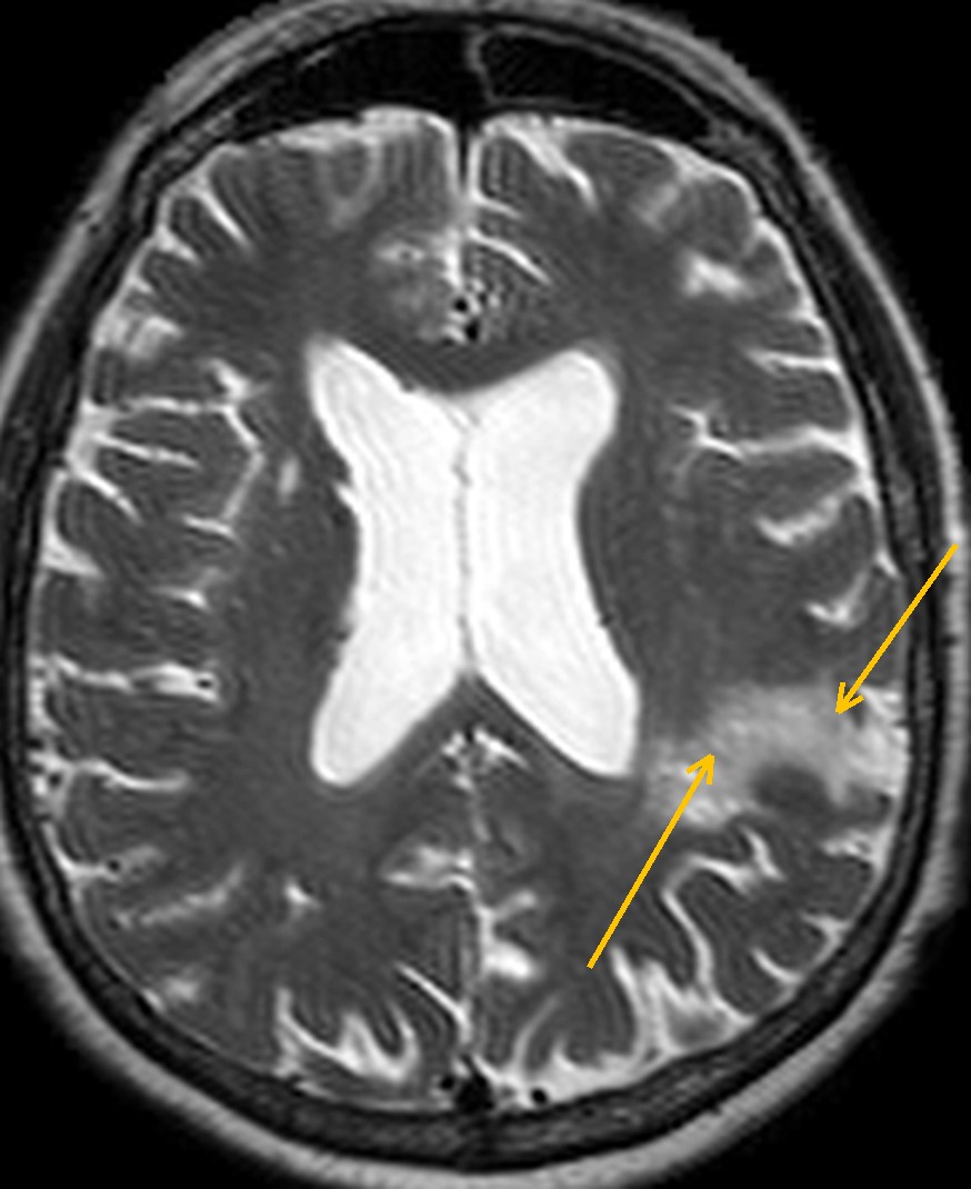
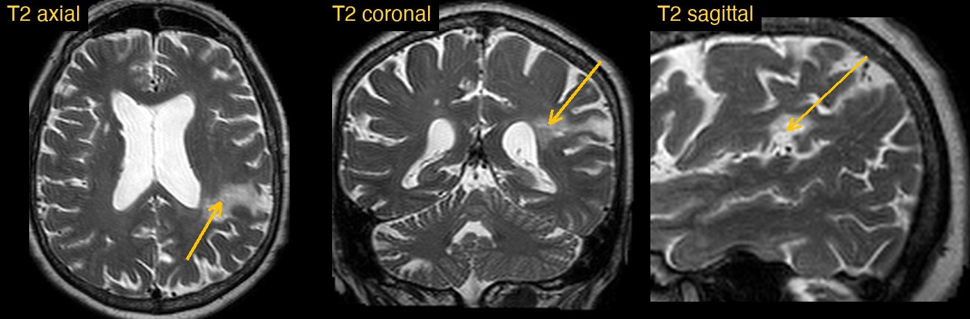
How Monitraq's AI read the scan
- It located the change: patchy T2 brightness in the cortex and in the subcortical and deep white matter around the back of the left Sylvian fissure, in the parietal operculum, posterior insula and perisylvian cortex, extending towards the lateral wall of the left lateral ventricle.
- It read the signs of tissue loss: there was no mass effect. The adjacent left Sylvian fissure was widened, and the left temporal horn and atrium were larger than the rest of the ventricles, which it read as ex vacuo change.
- It named the pattern: its impression favoured a chronic left posterior MCA branch infarct with encephalomalacia. It added that small bright foci high in the left parietal convexity might be further border-zone involvement.
- It kept the background separate: generalised volume loss with in-proportion ventricular enlargement and no features of hydrocephalus; a few small CSF-like deep white matter foci, most likely perivascular spaces, with chronic lacunes the main alternative; and a prominent CSF space beside the right cerebellum, possibly a small arachnoid cyst, with low confidence.
- It stated the limits: with T2 images only, acute ischaemia could not be excluded without DWI. It suggested DWI and ADC, FLAIR and SWI if clinically indicated, and carotid and intracranial vessel imaging if a vascular cause is confirmed.
The expert lesion mask that comes with this public dataset marks a chronic left MCA territory infarct of about 10 mL, centred posteriorly in the parietal and temporo-occipital region.
What is an MCA branch infarct?
The middle cerebral artery (MCA) runs out through the Sylvian fissure and divides into branches that fan over the surface of the frontal, parietal and temporal lobes. When the main trunk is blocked, a large part of the hemisphere can be lost. When only one branch is blocked, often by a small clot carried from the heart or a neck artery, the damage is a smaller wedge of cortex and the white matter beneath it.
Size does not equal importance. On the left side, the posterior perisylvian cortex is part of the language network in most people, so even a small infarct there can affect speech and understanding. Over weeks to months, dead tissue is cleared and replaced by fluid (encephalomalacia) and scar (gliosis), and the brain next to it draws back.
Key MRI signs of a chronic MCA branch infarct
- A wedge or patch of abnormal signal involving cortex and the white matter beneath it, within one MCA branch territory.
- Fluid-like signal in the core (encephalomalacia), with a bright rim of gliosis on FLAIR.
- Local volume loss: widened sulci or fissure and an enlarged nearby part of the ventricle (ex vacuo change).
- No mass effect and no restricted diffusion once the infarct is old.
- Sometimes dark spots on SWI from old blood products.
Differential diagnosis
A chronic infarct is usually recognised by its shape, its fit to an arterial territory and its volume loss. Old contusion from head injury tends to sit at the frontal and temporal poles. Border-zone infarcts lie between territories rather than within one. Demyelination favours the periventricular white matter and rarely shows cortical involvement on standard MRI. A low-grade tumour usually swells the cortex rather than shrinking it. The key question for the clinical team, old or new, needs DWI.
Frequently asked questions
How can MRI tell an old stroke from a new one?
Diffusion imaging is the key. A recent infarct is bright on DWI and dark on ADC, while an old one does not restrict diffusion and shows fluid, gliosis and volume loss instead.
Why is FLAIR useful in a chronic infarct?
FLAIR darkens free fluid, so a cavity looks dark and the gliotic rim around it stays bright. It also helps separate small chronic lacunes from perivascular spaces.
What does ex vacuo change mean on MRI?
It means a ventricle, fissure or sulcus has enlarged to fill space left by lost brain tissue. It is not caused by raised pressure, unlike hydrocephalus.
A draft for the radiologist
A small old infarct still matters: it can explain symptoms and it prompts a search for the cause. Monitraq's AI is built to find it, describe it plainly, keep background findings in proportion and say what the scan cannot show.
It gives a draft, not a diagnosis. The AI flags, describes and suggests. A radiologist confirms, corrects and signs.
Monitraq's AI produces draft reports to support qualified radiologists. It is not a diagnosis and is not intended for use by patients. All case details have been removed or generalised to protect patient privacy, and the images have been cropped and stripped of identifying information. Image: Aphasia Recovery Cohort (ARC), OpenNeuro, CC0.