Spinal cord compression from an epidural mass on MRI: how Monitraq's AI read it
Spinal cord compression on MRI is one of the few spine findings that cannot wait. In this whole-spine study, a soft-tissue mass filled the thoracic canal and squeezed the cord. Here is how Monitraq's AI read the scan, and why it put that finding at the very top of its draft.
MRI findings
The patient had a spine MRI with sagittal T2-weighted images of the cervical, thoracic and lumbar spine, plus sagittal T1 and axial T2 and T1 images of the thoracic spine. No contrast, STIR or diffusion images were part of the study.
In the mid-to-lower thoracic spine, the normal bright ring of cerebrospinal fluid (CSF) around the cord disappeared over several vertebral segments. In its place was soft tissue of intermediate signal, filling the canal and pushing on the cord. On the axial images the same tissue filled the canal and continued out to the left, beside the vertebral bodies.
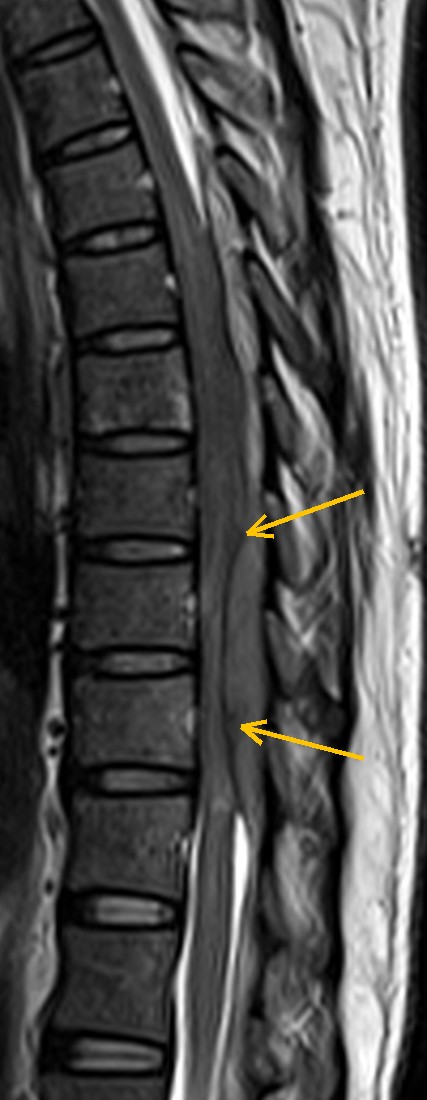
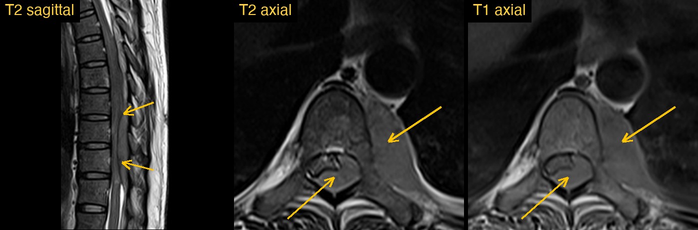
How Monitraq's AI read the scan
- It put the urgent finding first. The draft was triaged as urgent. Its first red flag was thoracic cord compression by a soft-tissue mass inside the canal, spanning several segments, with a block to the flow of CSF.
- It described the mass: a soft-tissue mass over about three to four vertebral segments of the mid-to-lower thoracic canal, intermediate on T2 and T1, with the cord compressed and displaced, no CSF around it on axial T2, and a capped, abruptly ending CSF column below it.
- It followed the tissue outside the spine. The mass continued through the left neural foramen into a left paravertebral, posterior mediastinal component lying along the vertebral bodies, a dumbbell shape.
- It checked the bones and discs: no vertebral collapse or disc destruction, and mildly patchy marrow signal next to the mass that it called indeterminate, since there was no STIR or contrast.
- It gave a differential: lymphoma was favoured, with a dumbbell nerve sheath tumour and tuberculous paravertebral or epidural disease as the main alternatives.
- It listed the next steps: same-day radiologist review; urgent contrast-enhanced thoracic MRI with STIR and diffusion and a whole-spine localiser to confirm the level count; CT of the chest, abdomen and pelvis to look for a primary tumour, lymph nodes or a tuberculous focus; urgent clinical assessment and spinal surgical or oncology review; and a biopsy of the paravertebral component as clinically appropriate.
Dr Priti, the radiologist who reviewed this case, reported spinal metastases: multiple vertebral marrow lesions with epidural and paravertebral soft tissue in the mid-lower thoracic spine compressing the cord.
The AI and the radiologist agreed on what mattered most that day: soft tissue in the epidural space and beside the spine, compressing the thoracic cord. They differed on the cause. That is exactly why a radiologist reviews and signs every report.
What is malignant spinal cord compression?
Malignant spinal cord compression happens when tumour tissue in or around the spine presses on the cord. Most often this is a metastasis in a vertebral body that grows backwards into the epidural space. Tumour can also reach the canal from the paravertebral tissues through the neural foramina, as lymphoma sometimes does.
The thoracic spine is the most common level. It is treated as an emergency, because function lost before treatment is often not regained, and MRI of the whole spine is the test of choice.
Key MRI signs of spinal cord compression from a tumour
- Loss of the CSF signal around the cord, with the cord flattened or displaced.
- Epidural soft tissue that is usually intermediate on T1 and T2 and enhances after contrast.
- A capped or abruptly ending CSF column above or below the mass on sagittal T2.
- Vertebral marrow lesions that replace the normal bright fatty marrow on T1, often bright on STIR.
- Paravertebral soft tissue, sometimes passing through a neural foramen in a dumbbell shape.
Differential diagnosis
- Metastases: usually start in the vertebral body or pedicle, with marrow replacement on T1 and often several levels involved.
- Lymphoma: can wrap around the spine and pass through the foramina with relatively little bone destruction.
- Nerve sheath tumour: a dumbbell mass that often widens the neural foramen and tends to grow slowly.
- Tuberculosis: usually involves the disc and the vertebral endplates, with paravertebral abscesses and spread under the ligaments.
Frequently asked questions
Which MRI sequences are best for spinal cord compression?
Sagittal T1 and T2 of the whole spine are the core. T1 shows marrow replacement well, T2 shows the cord and the CSF around it, and axial images and contrast show the epidural tissue and its extent.
Why image the whole spine?
Metastatic disease often affects more than one level, and a second area of compression can change treatment. A whole-spine view also helps to count the levels correctly.
What does a vertebral metastasis look like on T1?
It usually appears as a darker area that replaces the normal bright fatty marrow. Involvement of the pedicle or the back of the vertebral body raises concern for epidural spread.
A draft for the radiologist
In a spine study, order matters. The cord compression went first, with a clear note on what to do the same day. The details came after.
Monitraq's AI gives a draft, not a diagnosis. The AI flags, describes and suggests. A radiologist confirms, corrects and signs.
Monitraq's AI produces draft reports to support qualified radiologists. It is not a diagnosis and is not intended for use by patients. All case details have been removed or generalised to protect patient privacy, and the images have been cropped and stripped of identifying information.