Scattered bright dots after a road accident: how Monitraq's AI reads them
An adult had a brain MRI after a road traffic accident. A few tiny bright spots on one sequence pointed to a diagnosis that is easy to overlook: cerebral fat embolism. Here is how Monitraq's AI worked through the scan.
The scan
The patient had a brain MRI after a road traffic accident. There was no bleed or swelling and nothing pressing on the brain, so on a first look the scan seemed reassuring.
The clue was on diffusion-weighted imaging (DWI), the sequence that lights up brain tissue whose cells have recently been injured. Scattered through both sides of the brain were several tiny bright dots.

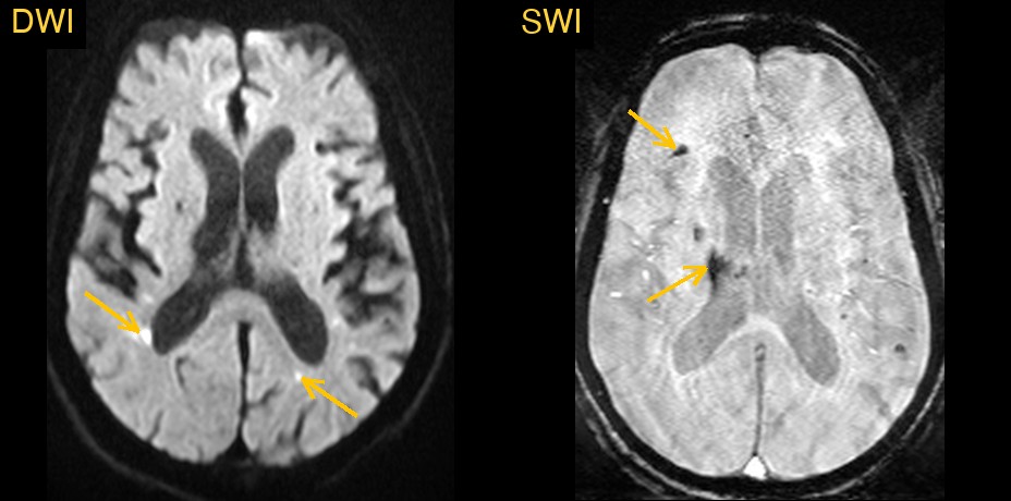
Why these dots are easy to overlook
This patient's brain also showed a lot of old change: extensive chronic small-vessel disease in the white matter, a few old lacunes and some age-related volume loss. In a brain like that, a few more small spots are easy to put down to "more of the same". That shortcut would miss what the accident may have caused.
How Monitraq's AI read the scan
- It described the dots precisely: multiple punctate and small DWI-bright foci in both hemispheres, at the grey-white junction and in the deep and periventricular white matter, seen on several neighbouring slices.
- It read the pattern: the dots were not in one artery's territory, which argues against a single stroke, and they sat separate from the old white matter damage.
- It put the history together with the images: new scattered DWI foci in a patient after a road accident made cerebral fat embolism the leading consideration. Diffuse axonal injury and embolic infarcts, including from a torn neck artery (dissection), came next.
- It weighed the other findings: small dark spots on susceptibility imaging (SWI) could be from the trauma or the fat embolism, not only old hypertensive microbleeds. The chronic small-vessel disease and volume loss were reported, but kept in the background.
- It flagged the scan for same-day radiologist review and listed the next steps: confirm true restricted diffusion on the ADC map, check for long-bone or pelvic fractures, low oxygen and a petechial rash, consider imaging the neck arteries if the embolic pattern was favoured, and consider a follow-up MRI.
Dr Priti, the radiologist who reviewed this case, also listed cerebral fat embolism among the possible causes of these foci.
What is cerebral fat embolism?
Fat embolism happens when fat droplets enter the bloodstream after a broken bone, most often a long bone such as the femur or the pelvis. They lodge in tiny brain vessels and cause a shower of small injuries, usually one to three days after the trauma. On DWI that shower can look like a sky full of stars, sometimes called the starfield pattern. Doctors also watch for low oxygen levels, confusion and a pinpoint rash on the chest or in the eyes.
Recognising it matters because it changes what the clinical team looks for next: fractures, oxygen levels and how the patient is changing hour by hour.
A draft for the radiologist
A good radiologist reads the images together with the story: this patient was just in an accident. Monitraq's AI is built to do the same, and to show its reasoning so the radiologist can check it quickly.
It gives a draft, not a diagnosis. The AI flags, describes and suggests. A radiologist confirms, corrects and signs.
Monitraq's AI produces draft reports to support qualified radiologists. It is not a diagnosis and is not intended for use by patients. All case details have been removed or generalised to protect patient privacy, and the image has been cropped and stripped of identifying information.