Swelling at the back of the brain: how Monitraq's AI reads PRES
An adult had a brain MRI that showed swelling in the back of both sides of the brain, with a smaller patch near the top of the frontal lobes and a few small bleeds. Where the swelling sits is the key to the diagnosis. Here is how Monitraq's AI worked through the scan.
The scan
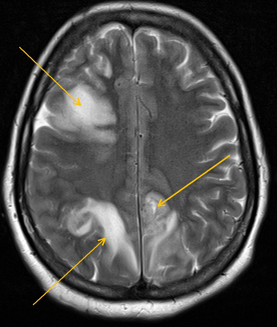
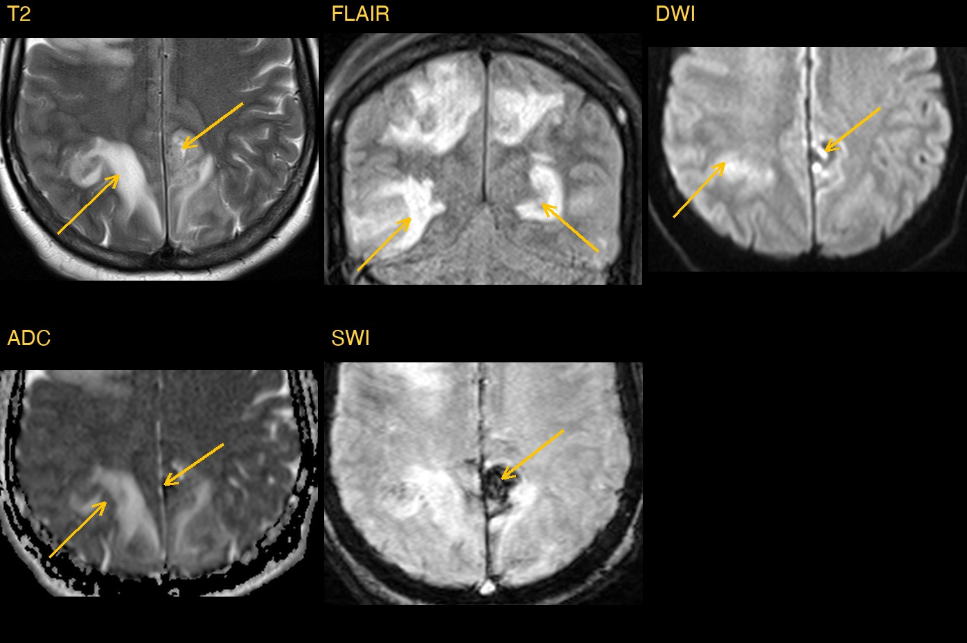
On T2 and FLAIR images, both parietal and occipital lobes were bright and swollen, in the cortex and in the white matter just beneath it. The change was on both sides and roughly symmetric. A similar, smaller area sat higher up in the frontal lobes, along the superior frontal sulcus.
Two other sequences added important detail. The ADC map showed that water was moving more freely than normal in the swollen areas, the sign of vasogenic oedema (fluid leaking out of blood vessels) rather than a stroke. And susceptibility imaging (SWI), which is very sensitive to blood, showed a small bleed in the left parietal lobe and several tiny bleeds inside the swollen areas.
Why the location matters
Bright white matter on T2 and FLAIR has many causes, including infection, inflammation, inherited white matter disease and stroke. What narrows the list here is the distribution: both sides, mostly at the back of the brain, in the cortex and the white matter just under it, with a band higher up along the frontal convexities. Add raised ADC and small bleeds, and one diagnosis stands out.
How Monitraq's AI read the scan
- It mapped the swelling: bilateral, roughly symmetric, confluent cortical and subcortical T2/FLAIR brightness with gyral swelling, centred on the parieto-occipital lobes and spreading to the high convexities, plus similar change in the right frontal lobe at the superior frontal sulcus and a smaller area on the left high frontal convexity.
- It checked the diffusion: ADC was clearly raised, so this was vasogenic oedema. Brightness on DWI in these areas was explained as T2 shine-through, not true restriction.
- It found the bleeding: a small parenchymal haematoma in the left medial parietal lobe, near the falx, with marked blooming on SWI, and several small punctate and curvilinear SWI-dark foci in both occipital lobes and the right frontal lobe, consistent with petechial haemorrhage.
- It named the pattern: posterior and superior frontal sulcus cortical-subcortical vasogenic oedema, typical of posterior reversible encephalopathy syndrome (PRES), with a haemorrhagic component. Cerebral venous sinus thrombosis was named as the main alternative that must be excluded.
- It flagged the scan as urgent because of the bleeding, and listed the next steps: review the venogram or obtain venography to exclude venous thrombosis; check for PRES triggers such as blood pressure, pregnancy or the postpartum period, kidney function and immunosuppressive or cytotoxic drugs; and repeat the MRI after treatment to confirm resolution.
Dr Priti, the radiologist who reviewed this case, reported posterior reversible encephalopathy syndrome (PRES): vasogenic oedema in the cortex and subcortical white matter of both parieto-occipital regions and along the superior frontal sulci, with a small left medial parietal haematoma and petechial haemorrhages.
What is PRES?
Posterior reversible encephalopathy syndrome is a form of brain swelling that happens when the brain can no longer keep its blood flow steady, often during a sudden rise in blood pressure. Fluid leaks from small vessels into the brain tissue. The back of the brain is affected most often. Common triggers include severe high blood pressure, eclampsia, kidney disease and some immunosuppressive and cancer drugs.
Patients often have headache, confusion, seizures or problems with vision. As the name says, the changes are often reversible when the cause is treated quickly. Bleeding occurs in some patients and makes prompt recognition even more important.
A draft for the radiologist
A good radiologist looks at where an abnormality sits, not only what it looks like, and checks for the complications that change urgency. Monitraq's AI is built to do the same, and to show its reasoning so the radiologist can check it quickly.
It gives a draft, not a diagnosis. The AI flags, describes and suggests. A radiologist confirms, corrects and signs.
Monitraq's AI produces draft reports to support qualified radiologists. It is not a diagnosis and is not intended for use by patients. All case details have been removed or generalised to protect patient privacy, and the image has been cropped and stripped of identifying information.