Hirayama disease on cervical spine MRI: how Monitraq's AI read it
Hirayama disease can hide on a routine cervical spine MRI taken with the neck straight. In this young adult the clues were subtle: a straightened neck and a flattened lower cord. Here is how Monitraq's AI read the scan, and why it asked for one more test.
MRI findings
The study covered the cervical spine with sagittal T2 and T1, coronal STIR, and axial gradient-echo and T1 images, together with brain T2 and DWI. All of it was taken in the neutral position. There was no flexion series.
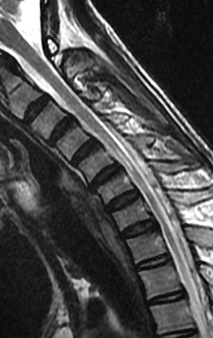
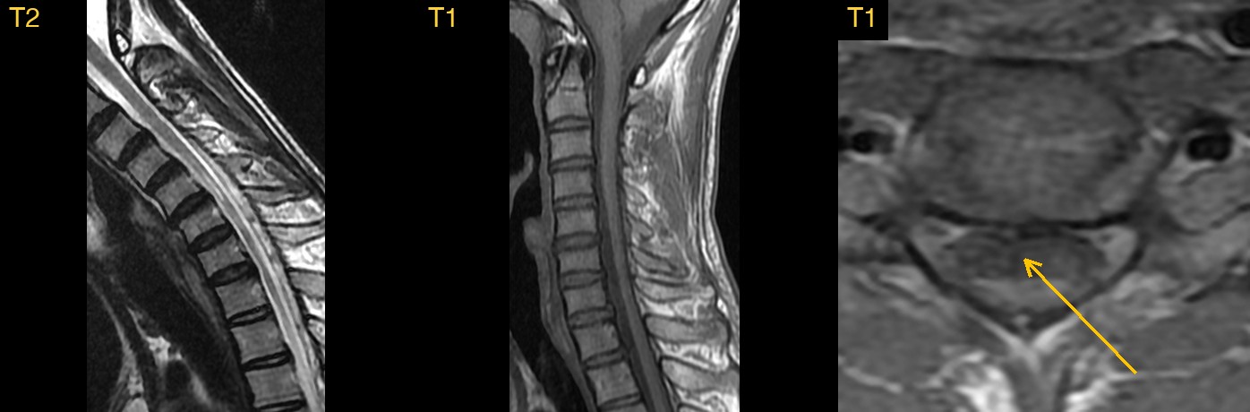
At first the scan looks almost normal: healthy discs, no narrowing of the canal and nothing pressing on the cord. The first clue is the shape of the neck. The normal gentle forward curve of the cervical spine (the lordosis) is lost, and the backs of the vertebral bodies form a straight line from C2 to C7.
The second clue is on the axial images. In the lower neck, the cord looks flattened from front to back and lies against the front of the canal, with more fluid behind it than usual.
How Monitraq's AI read the scan
- It described the alignment: loss of the cervical lordosis, with a straightened line along the backs of the vertebral bodies from C2 to C7 and no step-off.
- It described the cord shape: on the axial gradient-echo images the mid-lower cervical cord, about C5 to C7, appeared flattened front to back and lay against the front of the canal, with a widened space behind it. The cord was round at C1 and T1.
- It looked for a cause and found none: no disc herniation, canal or cord compression, listhesis, marrow lesion or flow voids. The brain images showed no acute or focal abnormality.
- It gave a ranked differential: Hirayama disease (juvenile monomelic amyotrophy) as the leading diagnosis, which a neutral-position study cannot exclude; a normal oval shape of the cervical enlargement with positional straightening as possible; and motor neuron disease or post-inflammatory cord atrophy as unlikely.
- It said what would settle it: a flexion cervical MRI with contrast, with sagittal and axial T2 and post-contrast T1 in full neck flexion, to look for forward shift of the dura and engorged veins behind it. It also suggested checking for asymmetric hand and forearm weakness or wasting, and nerve conduction studies with EMG.
Dr Priti, the radiologist who reviewed this case, reported Hirayama disease (juvenile asymmetric segmental spinal muscular atrophy, distal cervical myelopathy). She noted loss of the normal cervical lordosis on these neutral-position images and that a flexion MRI with contrast is needed to show the forward shift of the posterior dura and the posterior epidural venous plexus.
What is Hirayama disease?
Hirayama disease is a rare cause of weakness and wasting in the hand and forearm. It mainly affects young men in their teens and twenties. The weakness is usually on one side or clearly worse on one side. It tends to progress slowly for a few years and then stop.
The leading explanation is mechanical. When the neck bends forward, the back wall of the dural sac moves forward and presses the lower cervical cord against the vertebral bodies. Repeated over time, this is thought to injure the anterior horn cells that supply the hand muscles.
So neck position matters. In the neutral position the changes are subtle. In flexion, the forward shift of the dura can be seen directly.
Key MRI signs of Hirayama disease
- Loss of the normal cervical lordosis on neutral-position images.
- Flattening or thinning of the lower cervical cord, often asymmetric, around C5 to T1.
- Loss of the normal contact between the back of the dural sac and the bone behind it.
- Sometimes faint T2-bright signal in the lower cervical cord.
- On flexion: forward shift of the posterior dura and a crescent-shaped space behind it, with flow voids and enhancement from engorged epidural veins.
Differential diagnosis
- A normal cord with positional straightening. The cervical cord is normally oval, and a straight neck can be positional. Normal cord measurements and no change on flexion support this.
- Motor neuron disease. Weakness usually spreads beyond one limb, and EMG shows widespread denervation.
- Cervical spondylotic myelopathy. There is a compressive cause, such as a disc or bony ridge, usually in an older patient.
Frequently asked questions
Which MRI is needed to diagnose Hirayama disease?
A flexion MRI of the cervical spine, ideally with contrast. Sagittal and axial images in full neck flexion show the forward shift of the posterior dura and the engorged veins behind it.
Can a routine cervical spine MRI miss Hirayama disease?
Yes. Neutral-position images may show only loss of lordosis and subtle cord flattening, or look normal. Flexion imaging is the key test.
What does the flattened cord look like on axial images?
The lower cervical cord looks flat from front to back, sometimes more on one side, and lies against the front of the canal.
A draft for the radiologist
Here the AI linked two quiet findings to a named condition, said plainly that a neutral-position study could not exclude it, and set out the flexion study that would answer the question.
It gives a draft, not a diagnosis. The AI flags, describes and suggests. A radiologist confirms, corrects and signs.
Monitraq's AI produces draft reports to support qualified radiologists. It is not a diagnosis and is not intended for use by patients. All case details have been removed or generalised to protect patient privacy, and the images have been cropped and stripped of identifying information.