Papillary tumour of the pineal region on MRI: how Monitraq's AI read it
This brain MRI shows a papillary tumour of the pineal region: a lobulated solid-cystic mass behind the third ventricle, pressing on the aqueduct and causing obstructive hydrocephalus. Here is how Monitraq's AI worked through the scan.
MRI findings
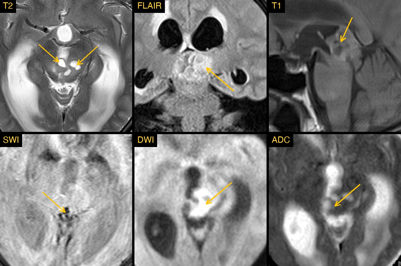
The study had T2, FLAIR, T1, susceptibility-weighted (SWI) and diffusion-weighted (DWI) images, with an ADC map. There was no contrast-enhanced series.
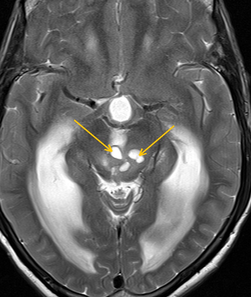
The first thing that stands out is the size of the ventricles. Both lateral ventricles and the third ventricle are ballooned, while the fourth ventricle is normal in size. That pattern points to a block at the cerebral aqueduct, the narrow channel between the third and fourth ventricles.
The cause sits just above it: a lobulated mass in the pineal region, behind the third ventricle and above the tectal plate. On T2 it contains several bright, fluid-like cysts within darker solid tissue.
How Monitraq's AI read the scan
- It described the mass: a single lobulated solid-cystic mass in the pineal region, posterior third ventricle and quadrigeminal cistern, about 25 x 22 mm on the midline sagittal image. It noted several T2-bright cysts, a heterogeneous solid part of intermediate T1 signal containing intrinsic T1-bright foci, foci of susceptibility on SWI, and patchy DWI brightness in the solid part without clear ADC darkening.
- It traced the effect on the ventricles: the mass compresses the tectal plate and aqueduct, causing obstructive triventricular hydrocephalus. It reported ballooned lateral ventricles, enlarged temporal horns, an enlarged third ventricle, upward bowing and thinning of the corpus callosum and a normal-sized fourth ventricle. It measured an Evans index of about 0.36, above the usual upper limit of 0.3.
- It recognised pressure on the ventricle walls: broad, confluent bright signal around the frontal horns, bodies and occipital horns on T2 and FLAIR, with raised ADC, which it read as CSF seeping across the ventricle lining (transependymal seepage).
- It gave a ranked differential: papillary tumour of the pineal region favoured, with pineal parenchymal tumour (pineocytoma, PPTID, pineoblastoma) and germ cell tumour (germinoma, teratoma) as alternatives, and a complex pineal cyst with haemorrhage unlikely because of the solid part and the hydrocephalus.
- It flagged the scan as urgent and listed next steps: urgent neurosurgical consultation for the hydrocephalus; contrast MRI of the brain with thin sagittal images through the pineal region and contrast MRI of the whole spine to look for spread in the CSF; serum AFP and beta-hCG; CSF markers and cytology only once neurosurgery judged it safe; and a non-contrast CT head to characterise calcification.
It also stated what the study could not show. Without contrast, enhancement of the mass and any spread along the ventricle lining or meninges could not be judged. Without SWI phase images, calcium could not be told apart from blood in the mass.
Dr Priti, the radiologist who reviewed this case, reported a papillary tumour of the pineal region causing obstructive hydrocephalus.
What is a papillary tumour of the pineal region?
A papillary tumour of the pineal region is a rare neuroepithelial tumour. It is thought to arise from specialised ependymal cells of the subcommissural organ, a small structure at the back of the third ventricle near the entrance to the aqueduct. It is seen in children and adults, most often in young adults.
The WHO classification grades it as grade 2 or 3. Diagnosis rests on histology. Local recurrence is a recognised problem, so long-term imaging follow-up is usual after treatment.
Like other pineal region masses, it often comes to attention through hydrocephalus, because the aqueduct lies directly beneath the pineal gland. Pressure on the tectal plate can also affect eye movements.
Key MRI signs of a papillary tumour of the pineal region
- A well-defined, lobulated mass in the pineal region or posterior third ventricle.
- Mixed solid and cystic parts, with the cysts bright on T2.
- Intrinsic T1-bright foci on the unenhanced T1 images, a described clue to this tumour.
- Heterogeneous enhancement of the solid part after contrast.
- Obstructive hydrocephalus of the lateral and third ventricles from compression of the aqueduct.
Differential diagnosis
A pineal region mass has a fairly short list of causes. Imaging narrows it, but rarely settles it.
- Pineal parenchymal tumours (pineocytoma, pineal parenchymal tumour of intermediate differentiation, pineoblastoma). Pineal calcification pushed outwards ("exploded") on CT is a classic clue. Marked restricted diffusion and CSF seeding favour pineoblastoma.
- Germ cell tumours, especially germinoma. These are more common in children and young men. Raised AFP or beta-hCG in blood or CSF helps, and teratomas may contain fat.
- Pineal cyst. A simple cyst follows CSF signal and has no solid nodule. A solid component or hydrocephalus argues against it.
- Other tumours such as meningioma of the tentorial region or a glioma of the tectal plate can also present here.
Frequently asked questions
Which MRI sequence best shows a papillary tumour of the pineal region?
T2 images show the solid and cystic parts and the hydrocephalus well. Unenhanced T1 matters too, because intrinsic T1-bright foci are a described clue. Contrast-enhanced T1, ideally with thin sagittal images, shows the enhancing solid part and helps look for spread.
Why does a pineal region mass cause hydrocephalus?
The cerebral aqueduct runs just beneath the pineal gland and tectal plate. A mass here can squeeze it shut, so CSF builds up in the lateral and third ventricles while the fourth ventricle stays normal in size.
Why image the whole spine?
Pineal region tumours can spread through the CSF and seed the spinal canal. A contrast-enhanced MRI of the whole spine is the usual way to look for this before treatment is planned.
A draft for the radiologist
In this case the urgent problem was the hydrocephalus, and the AI put that first. It then worked out the most likely type of tumour from the mix of cysts, T1-bright foci and location, and set out what still needed to be checked.
It gives a draft, not a diagnosis. The AI flags, describes and suggests. A radiologist confirms, corrects and signs.
Monitraq's AI produces draft reports to support qualified radiologists. It is not a diagnosis and is not intended for use by patients. All case details have been removed or generalised to protect patient privacy, and the images have been cropped and stripped of identifying information.