An old stroke on the left side of the brain: how Monitraq's AI reads it
An adult's brain MRI showed a large area on the left side where brain tissue had been replaced by fluid. The question was not whether something was there, but what it was and how old it was. Here is how Monitraq's AI worked through the scan.
The scan
This brain MRI comes from a public research collection. Only one type of image was available: a T2-weighted volume, on which water and fluid look bright.
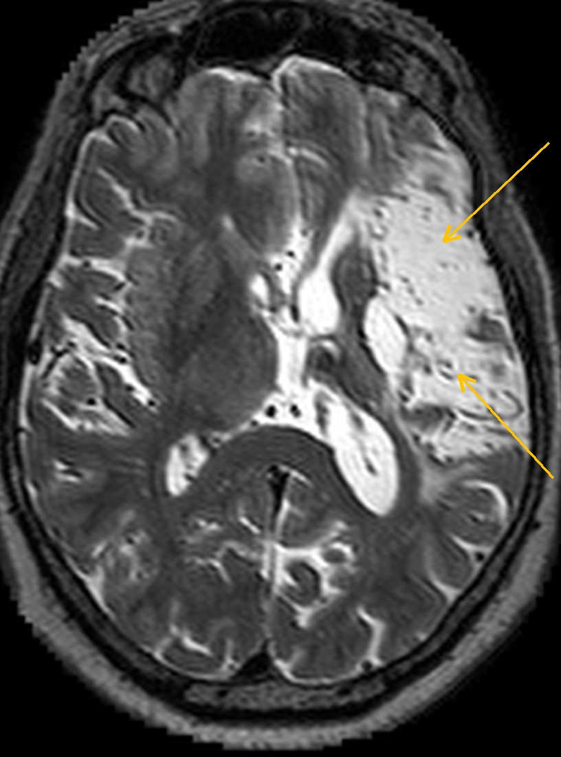
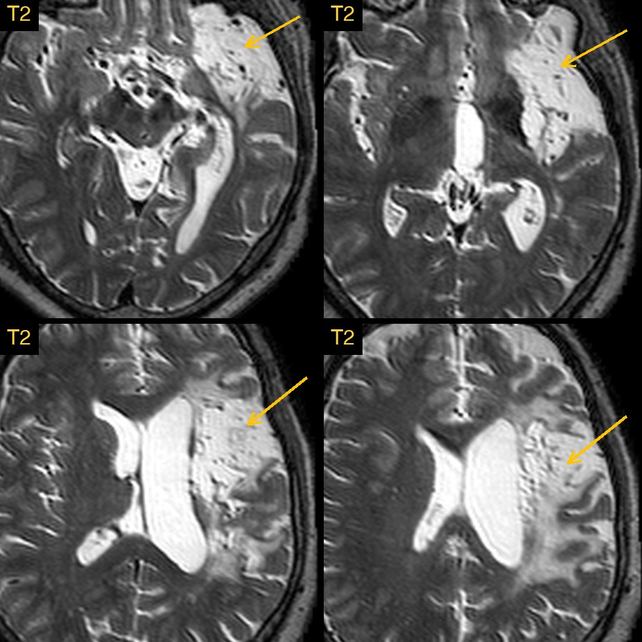
On the left side of the brain, around the insula and the lower part of the frontal lobe, a large region was as bright as the fluid in the ventricles. Normal brain tissue there had gone.
Why the age and the cause matter
A large bright area on T2 can mean very different things. A fresh stroke, a tumour, an old bleed or an old injury can all change the signal in this part of the brain. What the radiologist needs to say is whether this is old, settled damage or something active, and which pattern it fits. That decides whether the next step is routine secondary prevention or urgent further imaging.
How Monitraq's AI read the scan
- It described the area precisely: a large region of CSF-like signal replacing the left insular cortex, the frontal operculum and the anterior temporal lobe, reaching the inferior frontal lobe, the superior temporal lobe and the deep white matter next to the lentiform nucleus, with bright scarred (gliotic) margins.
- It read the signs of an old process: vessels still ran through the cavity, and nothing was being pushed aside: no mass effect, no midline shift and no collection outside the brain. Tissue that has broken down and been cleared away behaves like this; a growing lesion does not.
- It matched the pattern to an artery: the damage followed the territory of the left middle cerebral artery (MCA) rather than being scattered through the white matter. It concluded: a chronic left MCA territorial infarct with encephalomalacia, centred on the insula and frontal operculum.
- It named the alternative: an old bleed or an old traumatic cavity was less likely, and SWI would help tell them apart.
- It was clear about the limits: with no DWI, a new stroke elsewhere could not be excluded. It suggested DWI, ADC and FLAIR if there were new symptoms, SWI for blood products, and imaging of the left carotid and middle cerebral arteries (MRA or CTA) for secondary prevention, if not already done.
The expert lesion mask that comes with this public dataset marks a chronic left MCA territory infarct of about 127 mL.
What is a chronic MCA infarct?
The middle cerebral artery is the largest branch of the internal carotid artery. It supplies most of the outer surface of the frontal, parietal and temporal lobes, the insula, and through small deep branches the basal ganglia and internal capsule. When it or one of its main branches is blocked, a large wedge of brain can die.
Over the following weeks and months the dead tissue is cleared away. What is left is a fluid-filled cavity (encephalomalacia) with a rim of scar tissue (gliosis), and the nearby ventricle and fissures widen to fill the space. On the left side the MCA territory includes the main language areas in most people, so a left MCA stroke often affects speech and language as well as strength on the right side of the body.
A draft for the radiologist
A good radiologist does not stop at "there is a big abnormality". They say what it is, how old it looks, what it is not, and what the scan cannot show. Monitraq's AI is built to do the same, and to show its reasoning so the radiologist can check it quickly.
It gives a draft, not a diagnosis. The AI flags, describes and suggests. A radiologist confirms, corrects and signs.
Monitraq's AI produces draft reports to support qualified radiologists. It is not a diagnosis and is not intended for use by patients. All case details have been removed or generalised to protect patient privacy, and the image has been cropped and stripped of identifying information. Image: Aphasia Recovery Cohort (ARC), OpenNeuro, CC0.