L5-S1 disc degeneration on MRI: how Monitraq's AI graded it
L5-S1 disc degeneration on MRI is one of the commonest findings in a lumbar spine report, and Pfirrmann grading puts a number on it. Here is how Monitraq's AI graded a lumbar scan level by level, and why its grade and the expert grade sat one step apart.
MRI findings
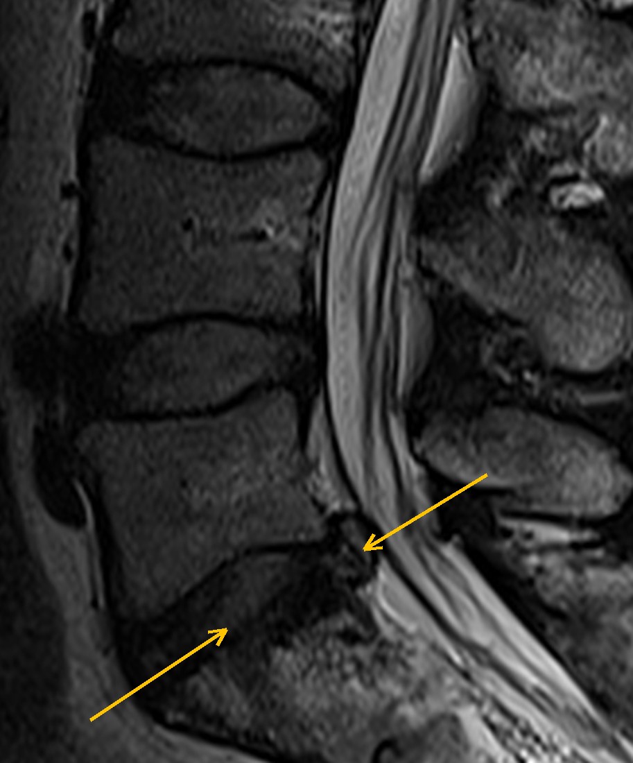
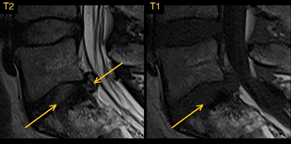
This lumbar spine MRI comes from a public research collection. The AI was given sagittal T1 and T2 images from about T11 to the sacrum. On T2, a healthy disc has a bright centre.
The AI read the lowest disc, L5-S1, as the most worn: lower than the discs above, dark on T2, with a small protrusion at its back edge. It found no narrowing of the spinal canal. One level up, it read the L4-L5 disc as greyer than normal, with a shallow bulge.
How Monitraq's AI read the scan
- It counted the levels upward from the sacrum, noting that transitional anatomy could not be excluded without a whole-spine view.
- It graded the L5-S1 disc: reduced disc height with diffuse loss of the T2 signal of the nucleus, Pfirrmann grade 4. It also read mild irregularity of the L5 and S1 endplates.
- It described the L5-S1 contour: a small posterocentral disc protrusion indenting the ventral epidural fat. Fluid around the descending sacral nerve roots was preserved, with no central canal stenosis. It raised possible mild narrowing of both L5-S1 foramina.
- It graded the L4-L5 disc: intermediate T2 signal in the nucleus with a prominent cleft, and preserved height, Pfirrmann grade 3. A shallow, diffuse posterior bulge only just indented the thecal sac.
- It checked the rest: alignment and vertebral heights were preserved, and the conus was normal at about L1.
- It set out the limits and next steps. With no axial images, the lateral recesses and foramina were only partly assessable. It suggested clinical correlation for L5 or S1 root symptoms and axial T2 from L3 to S1 if they persist, with no urgent imaging follow-up.
The expert gradings that come with this public dataset record, at L5-S1, mild disc degeneration (Pfirrmann grade 3) with disc space narrowing, a disc herniation and irregularity of both endplates; at L4-L5, Pfirrmann grade 3 with a diffuse disc bulge; and an overall impression of disc herniation at L5-S1.
The AI and the expert gradings agree on the shape of the case. L5-S1 is the most affected disc: narrowed, with irregular endplates and a small posterior herniation, which the AI called a protrusion, the commonest type of herniation. Both graded L4-L5 as Pfirrmann grade 3 with a bulge.
They differ on one point. The AI graded the L5-S1 disc one grade higher than the expert gradings: Pfirrmann 4 against Pfirrmann 3. Pfirrmann grading is a visual judgement, and grades 3 and 4 share a grey, poorly defined nucleus, so experienced readers often differ by one grade on the same disc. That is normal reader variation, and one reason a radiologist reviews and signs every report.
What is lumbar disc degeneration?
Each intervertebral disc has a soft, water-rich centre (the nucleus pulposus) inside a tough fibrous ring (the annulus fibrosus). With age and load, the nucleus loses water, the disc loses height, and the ring can bulge or tear. L4-L5 and L5-S1 are the discs most often affected. Degenerative change is very common, including in people with no back pain.
Key MRI signs of disc degeneration
- Loss of the bright T2 signal of the nucleus (disc desiccation).
- Loss of the clear border between nucleus and annulus.
- Reduced disc height.
- A broad disc bulge, or a focal herniation such as a protrusion.
- Irregularity of the endplates and signal change in the marrow next to them.
- Gas in the disc (vacuum phenomenon), dark on all sequences.
Pfirrmann grading of the lumbar discs
Pfirrmann grading uses sagittal T2 images and looks at three things: the disc's signal, the border between nucleus and annulus, and the disc height. Grade I is a bright, even disc of normal height. Grade II is still bright but uneven, sometimes with a dark horizontal band, and keeps a clear border. Grade III is grey and uneven, with an unclear border and normal or slightly reduced height. Grade IV is grey to dark, the border is lost, and the height is normal or moderately reduced. Grade V is black, with the disc space collapsed.
Grades III and IV overlap in look, so a report that also describes the signal and height in words shows why a grade was chosen.
Differential diagnosis
A low, dark L5-S1 disc with irregular endplates is usually degenerative. Discitis looks different: the disc is often bright on T2, with eroded endplates and marrow oedema, best seen on fat-suppressed images. A rudimentary disc at a transitional lumbosacral level can also look small, which is why level counting matters.
Frequently asked questions
Which MRI sequence is used for Pfirrmann grading?
Sagittal T2-weighted images. The grade is based on the disc's signal, the border between nucleus and annulus, and the disc height.
What is the difference between Pfirrmann grade 3 and grade 4?
Both have an uneven disc with an unclear or lost nucleus-annulus border. A grade 3 disc is grey with normal or slightly reduced height; a grade 4 disc is grey to dark with the border lost and the height normal or moderately reduced.
Is a disc protrusion the same as a disc herniation?
A protrusion is one type of herniation: a focal displacement of disc material whose base is wider than the distance it projects. A bulge is different: a broad extension of the disc over more than a quarter of its circumference.
A draft for the radiologist
A good lumbar report goes level by level, grades each disc and says what is and is not pressing on the nerves. Monitraq's AI is built to do the same and to show its reasoning, so the radiologist can check a grade quickly and adjust it where their judgement differs.
It gives a draft, not a diagnosis. The AI flags, describes and suggests. A radiologist confirms, corrects and signs.
Monitraq's AI produces draft reports to support qualified radiologists. It is not a diagnosis and is not intended for use by patients. All case details have been removed or generalised to protect patient privacy, and the images have been cropped and stripped of identifying information. Image: SPIDER lumbar spine MRI dataset, CC BY 4.0.