Large chronic MCA infarct on MRI: how Monitraq's AI read it
A large chronic MCA infarct on MRI leaves a recognisable footprint: a fluid-filled cavity, a rim of scar and a brain that has drawn back around the gap. This scan shows all three. Here is how Monitraq's AI read them.
MRI findings
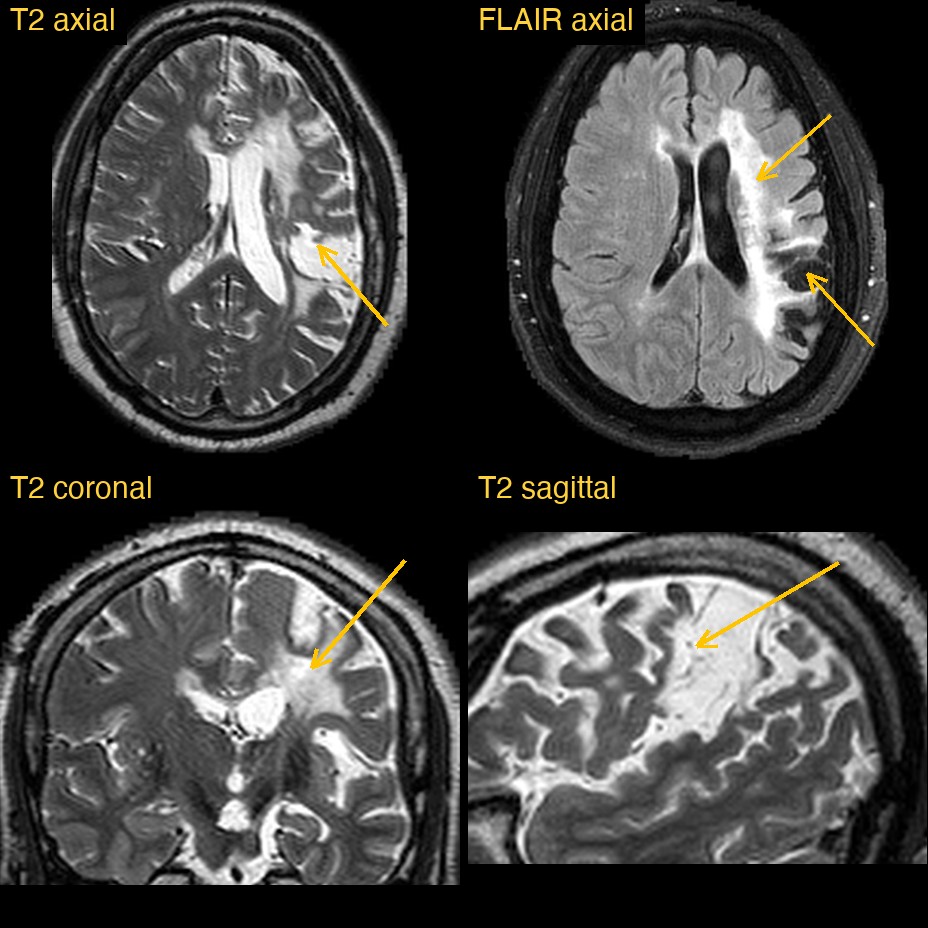
This brain MRI comes from a public research collection. It included a T2-weighted volume, viewed in three planes, and axial FLAIR images. On FLAIR, free fluid is dark, so scarred tissue stands out as bright.
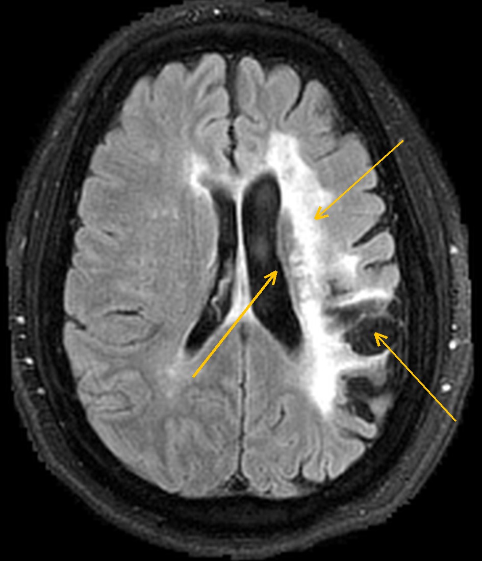
Over the left frontal and parietal lobes and around the left Sylvian fissure, a large area of brain had been replaced. On T2 it was as bright as the fluid in the ventricles. On FLAIR the same area was dark, edged by bright tissue that spread inwards to the wall of the left lateral ventricle. The left ventricle was larger than the right, and nothing was being pushed aside.
How Monitraq's AI read the scan
- It mapped the damage: a large region of CSF-signal cavitation in the left MCA territory, involving the posterior frontal (pre- and perirolandic), parietal and perisylvian and insular opercular cortex and the corona radiata and centrum semiovale beneath them.
- It used two sequences together: the cavity was bright on T2 and dark on FLAIR, like fluid, with a FLAIR-bright gliotic rim and confluent gliosis reaching the periventricular white matter.
- It read the volume loss: the nearby sulci were widened and the body and atrium of the left lateral ventricle were enlarged by ex vacuo change. There was no mass effect, hydrocephalus or collection outside the brain.
- It followed the consequences downstream: the left cerebral peduncle looked possibly smaller than the right, consistent with Wallerian degeneration of the left corticospinal tract. It noted that this was subtle.
- It kept the other hemisphere in view: patchy and partly chain-like T2/FLAIR bright foci in the right deep white matter, about Fazekas grade 2, favoured to be chronic small-vessel change, with internal border-zone ischaemia as the main alternative.
- It stated what the scan could not show: without DWI and SWI, a new infarct or old blood products could not be assessed. It suggested DWI and ADC if there are new symptoms, SWI for microbleeds, and carotid and intracranial vessel imaging.
The expert lesion mask that comes with this public dataset marks a chronic left MCA territory infarct of about 75 mL, centred on the frontoparietal and perisylvian region.
What is a chronic MCA infarct?
When a large branch of the middle cerebral artery is blocked, the brain it supplies dies. Over the following weeks and months, that tissue is broken down and cleared away. Three things are left behind. The first is encephalomalacia: a soft, fluid-filled gap where brain used to be. The second is gliosis: scar tissue formed by glial cells around and between the damaged areas. The third is ex vacuo change: the ventricle, sulci and fissures next to the gap widen to fill the space.
The damage can also travel. Nerve fibres that started in the dead cortex degenerate along their length, a process called Wallerian degeneration. In the corticospinal tract this can show as a smaller cerebral peduncle and pons on the same side.
Key MRI signs of a chronic MCA infarct
- A cavity in an MCA territory that follows fluid on every sequence: bright on T2, dark on FLAIR and T1.
- A bright rim of gliosis around the cavity on FLAIR.
- Widened sulci and fissures and an enlarged nearby ventricle, without mass effect.
- No restricted diffusion once the infarct is old.
- Sometimes Wallerian degeneration: a shrunken cerebral peduncle on the same side.
Differential diagnosis
A large fluid-filled area with gliosis is most often an old infarct when it fits an arterial territory, as here. An old haemorrhage leaves a cavity lined by dark haemosiderin on SWI. An old contusion favours the frontal and temporal poles. A porencephalic cavity from injury very early in life tends to have smooth walls, communicates with the ventricle and shows little gliosis. A tumour usually causes mass effect rather than volume loss.
Frequently asked questions
Why is an old infarct bright on T2 but dark on FLAIR?
The cavity is filled with fluid much like CSF. T2 shows that fluid as bright, while FLAIR suppresses it. Gliotic tissue holds water differently and stays bright on FLAIR.
What does Wallerian degeneration look like on MRI?
In the chronic stage, the affected tract shrinks and may show T2 brightness along its course. After a large MCA stroke this is usually seen as a smaller cerebral peduncle and pons on the side of the infarct.
How is ex vacuo ventricular enlargement different from hydrocephalus?
In ex vacuo change, the ventricle enlarges in step with lost brain and the nearby sulci are wide. In hydrocephalus the ventricles are out of proportion to the sulci, and there may be fluid seeping into the surrounding white matter.
A draft for the radiologist
An old stroke is more than "a big bright area". The report needs to say which territory it fills, what it has done to the rest of the brain and what the scan cannot rule out. Monitraq's AI is built to do that and show its reasoning.
It gives a draft, not a diagnosis. The AI flags, describes and suggests. A radiologist confirms, corrects and signs.
Monitraq's AI produces draft reports to support qualified radiologists. It is not a diagnosis and is not intended for use by patients. All case details have been removed or generalised to protect patient privacy, and the images have been cropped and stripped of identifying information. Image: Aphasia Recovery Cohort (ARC), OpenNeuro, CC0.