Tuberculous meningitis with hydrocephalus and ventriculitis on MRI: how Monitraq's AI read it
Tuberculous meningitis on MRI often shows itself through its effects: enlarged ventricles, a bright rim around them and material in the cisterns at the base of the brain. In this case all three were there. Here is how Monitraq's AI read the scan, and why the radiologist's final report matters.
MRI findings
The patient had a brain MRI with axial T2, coronal FLAIR, SWI, diffusion imaging with an ADC map, T1 images before and after contrast, FLAIR after contrast and MR spectroscopy.
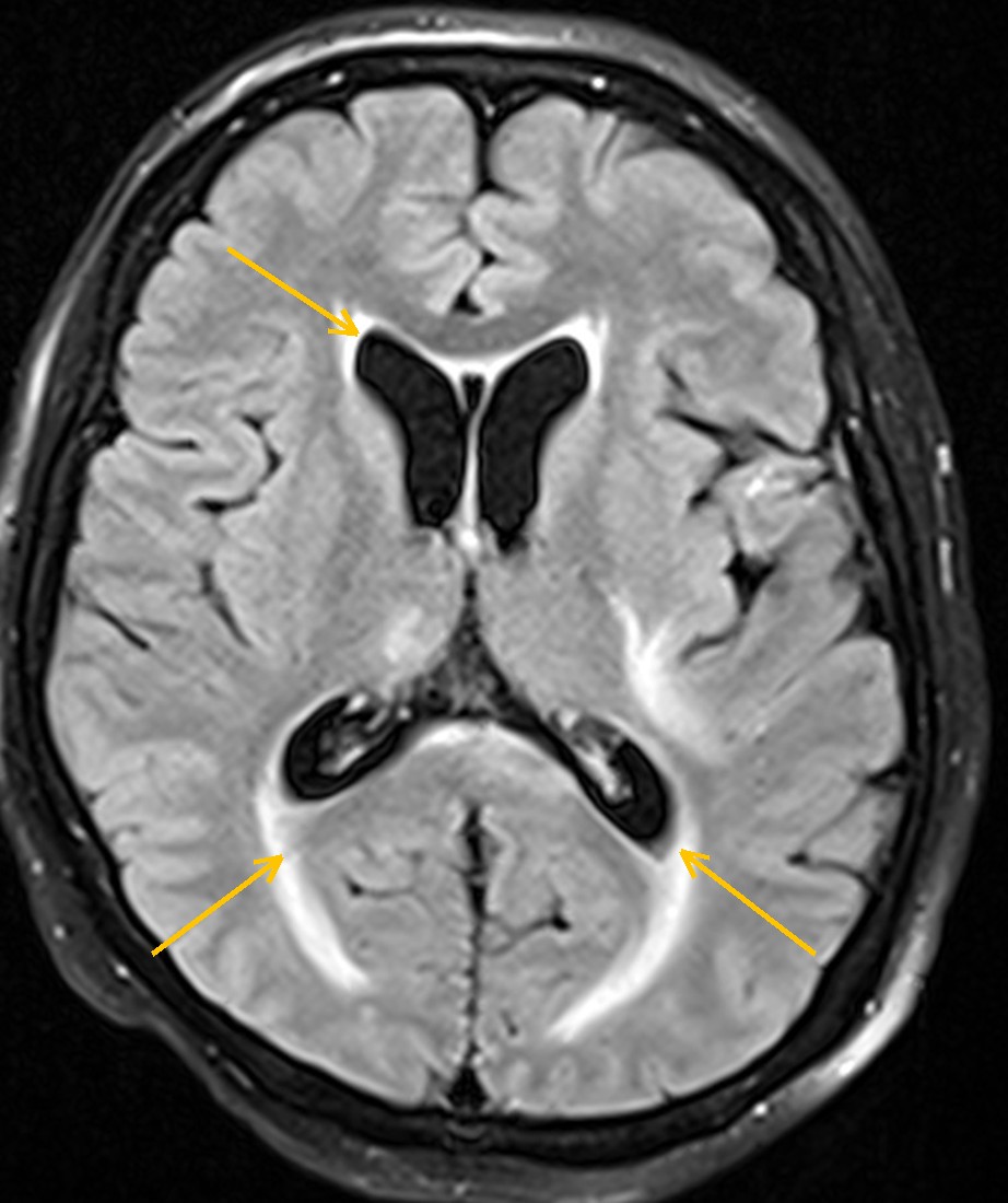
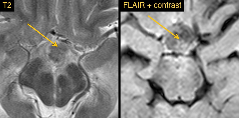
The lateral ventricles were enlarged, including the temporal horns, while the sulci over the brain were not widened to match. A smooth, bright rim on FLAIR surrounded the ventricles. Inside the back of the lateral ventricles there was material that was very bright on diffusion imaging. At the base of the brain, the cistern above the pituitary fossa did not hold clear fluid but soft material instead.
How Monitraq's AI read the scan
- It called hydrocephalus. The lateral ventricles were enlarged with rounded frontal horns and clearly dilated temporal horns, about 5 mm high against a normal of under about 2 mm. The sulci were not widened in proportion, which favours hydrocephalus over brain volume loss.
- It read the rim: a smooth, continuous FLAIR-bright rim around the lateral ventricles and temporal horns. With the enlarged ventricles, it described this as fluid seeping into the brain around the ventricles, or inflammation of the ventricle lining (ependymitis).
- It noted material inside the ventricles: very bright signal on diffusion imaging in the back of the lateral ventricles, left more than right.
- It put these together: hydrocephalus with probable ventriculitis, pyogenic or tuberculous. For the material in the ventricles, the main alternative was bleeding into the ventricles or a lesion of the splenium.
- It looked at the basal cisterns: the suprasellar and interpeduncular cisterns held intermediate T2 and bright post-contrast FLAIR material instead of clear fluid, with curved brightness along the left ambient cistern. It reported possible basal exudate at low confidence.
- It flagged the scan as urgent and listed the next steps: same-day radiologist review of the diffusion images; review of the post-contrast T1 for enhancement of the ventricle lining and the basal meninges; urgent clinical and CSF correlation for ventriculitis or meningitis, including a TB work-up; and a sagittal T2 to check the cerebral aqueduct.
Dr Priti, the radiologist who reviewed this case, reported a sellar tuberculoma: a lobulated, T2-hypointense, enhancing sellar and suprasellar lesion, with TB basal meningitis, hydrocephalus and ventriculitis.
The AI and the radiologist agreed on hydrocephalus and ventriculitis; the AI also flagged possible exudate in the suprasellar cistern at low confidence and asked for a TB work-up. The radiologist named the lesion as a tuberculoma and the cause as tuberculosis. That final diagnosis is why a radiologist signs every report.
What is tuberculous meningitis?
Tuberculous meningitis is infection of the meninges by Mycobacterium tuberculosis. It favours the base of the brain, where a thick exudate collects in the cisterns around the brainstem and the optic chiasm. It often develops over days to weeks rather than hours.
The exudate can block the flow of CSF and cause hydrocephalus. It can also inflame the small arteries at the base of the brain and cause infarcts. Some patients develop tuberculomas, which are firm, rounded collections of granulomatous tissue. Diagnosis rests on the clinical picture and CSF tests, with MRI showing the extent of disease and its complications.
Key MRI signs of tuberculous meningitis
- Thick enhancement of the basal meninges and exudate filling the basal cisterns.
- Hydrocephalus, with enlarged temporal horns as an early sign.
- A bright rim around the ventricles on FLAIR, from CSF seepage or ependymitis.
- Ependymal enhancement and debris in the ventricles in ventriculitis.
- Infarcts in the basal ganglia and thalami.
- Tuberculomas, often dark on T2 with ring or solid enhancement.
Differential diagnosis
- Bacterial (pyogenic) meningitis and ventriculitis: usually more acute; debris in the ventricles often restricts diffusion strongly.
- Fungal meningitis: can also favour the basal cisterns, especially in people with weak immunity.
- Neurosarcoidosis: basal meningeal and pituitary stalk involvement.
- Spread of cancer to the meninges: usually with a known primary tumour.
- Sellar masses: such as pituitary adenoma or craniopharyngioma, for a lesion in the sellar region.
Frequently asked questions
Which MRI sequence best shows tuberculous meningitis?
Post-contrast images show the basal meningeal enhancement best. Post-contrast FLAIR can make meningeal disease easier to see, as normal CSF stays dark.
What does a tuberculoma look like on MRI?
A tuberculoma is often dark on T2 because of its solid, caseating centre, with ring or solid enhancement. Some show a bright centre if they liquefy.
How does ventriculitis show on MRI?
Look for enhancement of the ventricle lining, debris in the dependent parts of the ventricles that is bright on DWI, and bright signal around the ventricles on FLAIR.
A draft for the radiologist
Enlarged ventricles, a bright rim and filled basal cisterns form a pattern that calls for an infection work-up. Monitraq's AI put that pattern together, raised tuberculosis as a possible cause and listed the tests to confirm it.
It gives a draft, not a diagnosis. The AI flags, describes and suggests. A radiologist confirms, corrects and signs.
Monitraq's AI produces draft reports to support qualified radiologists. It is not a diagnosis and is not intended for use by patients. All case details have been removed or generalised to protect patient privacy, and the images have been cropped and stripped of identifying information.