Nothing to find: how Monitraq's AI reads a normal brain MRI
A healthy volunteer had a research brain MRI. The hardest part of reading a normal scan is saying so clearly, without inventing findings and without hiding what the scan cannot show. Here is how Monitraq's AI worked through it.
The scan
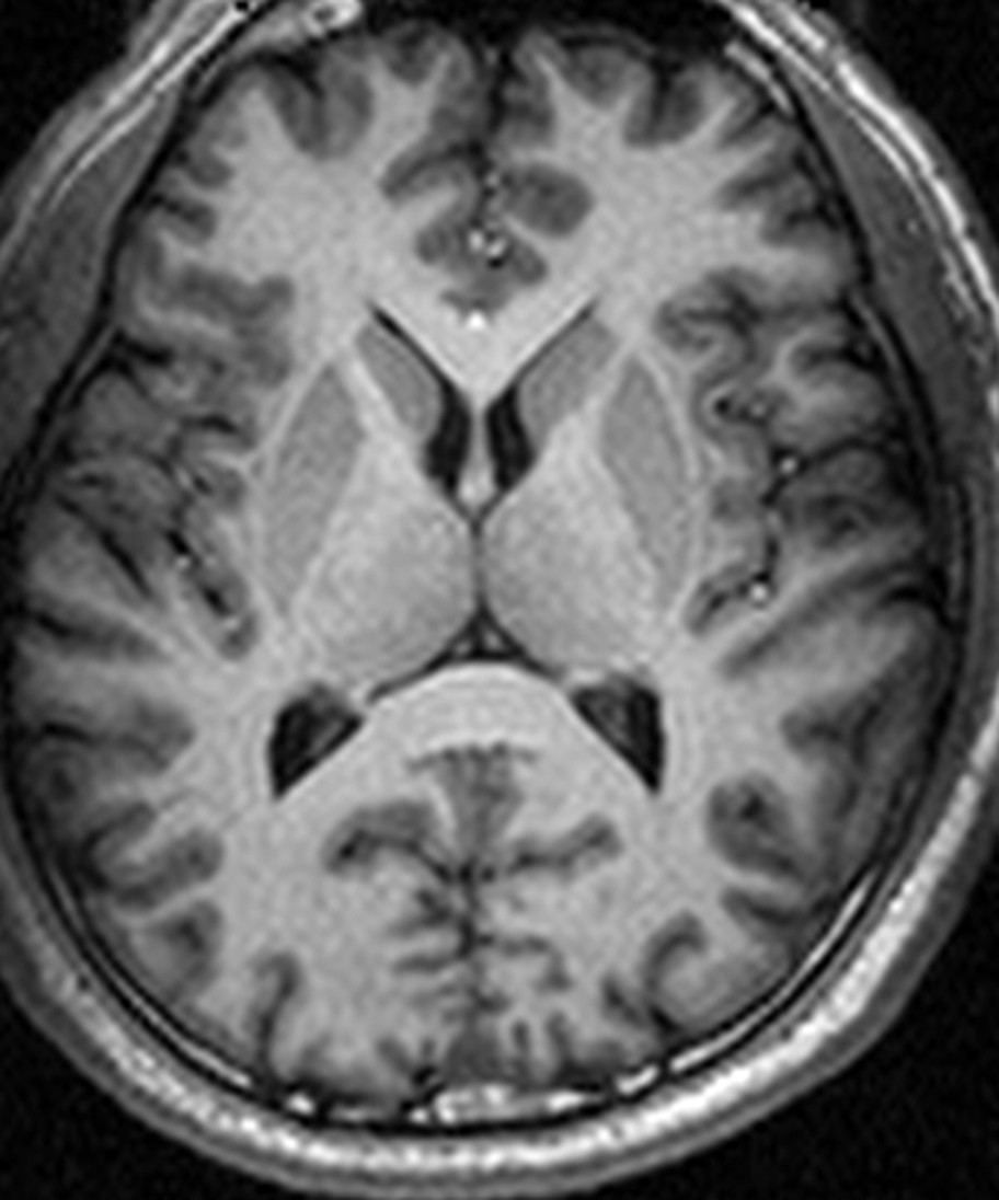
This was a research scan from a public dataset of healthy volunteers. It had a single sequence: a three-dimensional (volumetric) T1-weighted acquisition, which shows brain anatomy well. The face had been removed from the images by the dataset providers to protect the volunteer's identity.
There was no T2, FLAIR, diffusion or susceptibility imaging, and no contrast series.
Why a normal scan is not an easy read
On a detailed brain scan there is always something that can look unusual at first glance: a bright vessel, a small fluid space, a slight difference between the two sides. Calling these abnormal leads to worry, more tests and follow-up scans that were never needed. Calling a scan normal without saying what was not examined is just as unhelpful. A good normal report does both jobs: it says clearly that nothing is wrong, and it says what the scan could not show.
How Monitraq's AI read the scan
- It said the brain was normal, plainly: a normal-appearing brain, with no mass, haemorrhage, hydrocephalus, focal atrophy or structural anomaly on the sampled slices.
- It explained a finding that could have been over-called: the blood vessels in the head and neck looked bright. Rather than reading this as a sign that contrast had been given, or as something abnormal, it explained it as the normal effect of blood flowing into the slice on this type of T1 sequence, and noted there was no convincing evidence of contrast.
- It was honest about the limits: with T1 images only, it stated that acute stroke, white matter lesions, swelling and blood products could not be assessed, and that the face, orbits and sinuses were largely hidden by the dataset's defacing.
- It kept the next steps proportionate: review the full T1 volume to confirm there are no small lesions between the sampled slices. It added that T2, FLAIR, diffusion and susceptibility imaging would be needed only if clinically indicated, and are not required for a research or volunteer scan.
The dataset's reference reading for this scan is a normal brain MRI.
What does "normal" mean on a brain MRI?
A normal MRI report means that nothing abnormal was seen on the sequences that were done. It does not mean every possible problem has been ruled out: some conditions only show on particular sequences, such as diffusion imaging for a recent stroke or susceptibility imaging for tiny bleeds.
Small incidental findings are common on brain MRI, even in healthy people, and most of them do not matter. Telling them apart from real disease, and not reporting normal variants as problems, is part of reading a scan well.
A draft for the radiologist
A good radiologist is as careful with a normal scan as with an abnormal one: say it is normal, and say what it cannot show. Monitraq's AI is built to do the same, and to show its reasoning so the radiologist can check it quickly.
It gives a draft, not a diagnosis. The AI flags, describes and suggests. A radiologist confirms, corrects and signs.
Monitraq's AI produces draft reports to support qualified radiologists. It is not a diagnosis and is not intended for use by patients. All case details have been removed or generalised to protect privacy, and the image has been cropped and stripped of identifying information. Image: public healthy-volunteer dataset, OpenNeuro, CC0 (defaced by the dataset providers).