Plexiform neurofibroma of the face and neck on MRI: how Monitraq's AI read it
Plexiform neurofibroma on MRI can look like a cluster of bright nodules that ignores the normal boundaries of the face and neck. This case shows a patient with a large lesion of the left face and upper neck. Here is how Monitraq's AI read it, and where the radiologist's final answer came in.
MRI findings
The study was a head, face and neck MRI. It included coronal STIR and T1 images, axial and sagittal images, post-contrast T1 images and an MR angiogram of the neck.
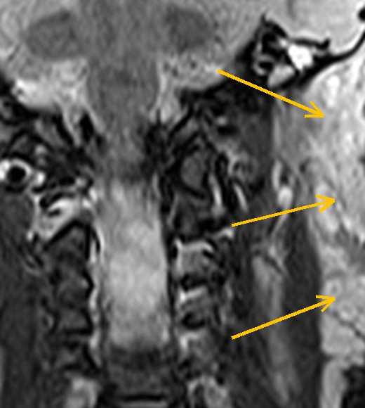
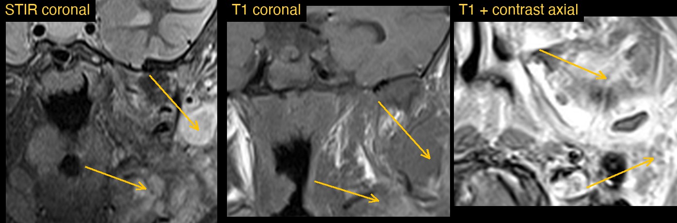
On the left, a large lobulated lesion spread through several deep spaces of the face and upper neck at once. It was made of many rounded, bright nodules on STIR, separated by strands of fat. The left side of the face was enlarged compared with the right. The right side looked normal.
How Monitraq's AI read the scan
- It mapped the extent. The lesion involved the left buccal and masticator regions, the left parotid region down to the angle of the jaw, the submandibular region and the left floor of the mouth and tongue. It reached the left retromandibular region and the upper side of the neck.
- It described the make-up: a large, ill-defined, trans-spatial, lobulated lesion made of many rounded STIR-bright nodules and channels with fat in between. It was intermediate on T1 and enhanced heterogeneously and avidly after contrast.
- It sized it roughly: about 9 cm from top to bottom on coronal STIR and about 4 cm thick across, with margins too ill-defined for an exact figure.
- It gave a differential: a low-flow vascular malformation (venous or venolymphatic) was favoured, with plexiform neurofibroma as the main alternative. A high-flow malformation or an aggressive tumour was not excluded on these images.
- It said how to separate them: look on the full series for phleboliths, fluid-fluid levels, flow voids and target signs, and consider a time-resolved contrast MR angiogram or Doppler ultrasound to tell low flow from high flow.
- It asked for the clinical picture: check for signs of neurofibromatosis type 1 (NF1), and refer to a vascular anomalies or head and neck team, with tissue sampling if the features were atypical.
Dr Priti, the radiologist who reviewed this case, reported a plexiform neurofibroma (NF1): a lobulated, trans-spatial, T2-bright nodular lesion of the left face and upper neck along the courses of nerves.
The AI and the radiologist described the same lesion: lobulated, bright on fluid-sensitive images and crossing spaces in the left face and neck. The AI listed plexiform neurofibroma as its main alternative. Weighing those two is the radiologist's job, which is why a radiologist signs every report.
What is a plexiform neurofibroma?
A plexiform neurofibroma is a benign tumour of the nerve sheath that grows along a nerve and its branches. It involves many nerve bundles at once, so it forms a tangle of thickened, rope-like strands. It is a hallmark of neurofibromatosis type 1.
In the head and neck it often follows branches of the trigeminal nerve or the nerves of the neck. It can cause facial asymmetry and overgrowth of the soft tissues. It usually shows in childhood. A small number turn malignant, so new pain or rapid growth needs prompt review.
Key MRI signs of plexiform neurofibroma
- A lobulated or multinodular mass that follows the course of nerves, sometimes called a "bag of worms".
- Spread across several spaces of the face or neck, often with no clear edge.
- Bright on T2 and STIR, often with a target sign: a darker centre and a brighter rim.
- Intermediate or low signal on T1.
- Variable, often heterogeneous enhancement after contrast.
- Widening of a skull base foramen or nerve canal when the tumour follows a nerve inwards.
Differential diagnosis
- Venous malformation: lobulated and very bright on T2, with dark round phleboliths and slow, gradual filling with contrast.
- Lymphatic malformation: fluid-filled cysts, often with fluid-fluid levels and only rim or septal enhancement.
- Arteriovenous malformation: flow voids and early filling on a time-resolved angiogram.
- Malignant peripheral nerve sheath tumour: a fast-growing, more heterogeneous part of a known plexiform neurofibroma.
Frequently asked questions
Which MRI sequence shows a plexiform neurofibroma best?
Fat-suppressed T2 or STIR images show it best. The nodules are bright against dark, suppressed fat, which shows the full extent and the target sign.
What is the target sign on MRI?
It is a nodule with a darker centre and a brighter rim on T2-weighted images. It is typical of neurofibromas and is less common in malignant nerve sheath tumours.
How can a venous malformation be told apart?
Phleboliths, which show as dark round dots, and slow, patchy filling on dynamic contrast images point to a venous malformation. Nodules along nerves, target signs and features of NF1 point to a plexiform neurofibroma.
A draft for the radiologist
A trans-spatial lesion in the face has a short list of causes, and several look alike. Monitraq's AI described the lesion, named the leading options and said which signs and tests would separate them.
It gives a draft, not a diagnosis. The AI flags, describes and suggests. A radiologist confirms, corrects and signs.
Monitraq's AI produces draft reports to support qualified radiologists. It is not a diagnosis and is not intended for use by patients. All case details have been removed or generalised to protect patient privacy, and the images have been cropped and stripped of identifying information.