Multiple small acute infarcts on MRI: how Monitraq's AI read them
Multiple small acute infarcts on MRI can be only a few millimetres across, and easy to lose among older white matter changes. In this adult's brain MRI, small bright spots on diffusion imaging sat in two different parts of the brain. Here is how Monitraq's AI read the scan.
MRI findings
The study included diffusion-weighted imaging (DWI) with an ADC map, axial FLAIR, susceptibility-weighted imaging (SWI), coronal T2 and a post-contrast T1 series.
The brain showed a lot of old change: many small bright spots in the white matter on FLAIR, an old small infarct on the right and some general volume loss. Among this, two new things stood out on DWI. One was a small bright focus in the deep white matter of the left cerebral hemisphere. The other was a bright area in the left posterior fossa, beside the fourth ventricle, where the brainstem joins the cerebellum.
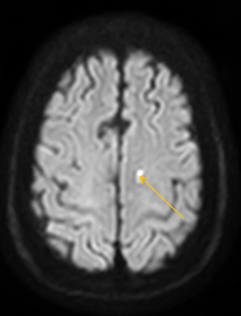
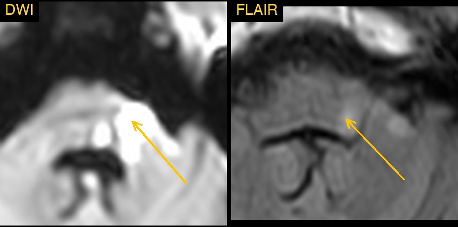
How Monitraq's AI read the scan
- It found the posterior fossa infarct: an area that was bright on FLAIR, with restricted diffusion and matching low signal on the ADC map, in the left posterior fossa where the brainstem joins the cerebellum.
- It found the second infarct: a separate small focus of restricted diffusion, also bright on FLAIR, in the deep white matter of the left cerebral hemisphere.
- It read the pattern: the two infarcts lay in different arterial territories, one in the back of the brain and one in the left hemisphere. Infarcts in more than one territory raise the question of an embolic source.
- It kept the old and the new apart. The many white matter spots on FLAIR did not restrict diffusion, so it reported them as chronic small-vessel change, along with an old small infarct on the right and age-related volume loss.
- It flagged the scan as urgent and listed the next steps: urgent stroke team review with the time of onset and the neurological findings; CT or MR angiography of the head and neck to look at the vertebral, basilar and carotid arteries; heart tests for a source of emboli, such as ECG or Holter monitoring and echocardiography; and assessment of blood pressure and vascular risk factors.
Dr Priti, the radiologist who reviewed this case, reported multiple tiny T2/FLAIR-bright foci with diffusion restriction in the left corona radiata, the left lateral medulla and the anterior lobe of the left cerebellum: acute lacunar infarcts, with a lateral medullary (Wallenberg) syndrome.
The AI and the radiologist agreed on the core of the case: small acute infarcts in the left deep white matter and the left posterior fossa. The radiologist's report gives the exact sites and the clinical syndrome. That final, precise word is why a radiologist signs every report.
What are multiple small acute infarcts?
An infarct is an area of brain that has died because its blood supply was cut off. Small acute infarcts can come from disease of a single tiny artery deep in the brain, or from small clots that travel from the heart or from a larger artery upstream.
When small infarcts appear in more than one arterial territory at the same time, an embolic cause becomes more likely. This changes the work-up: the heart and the neck arteries are checked, as well as blood pressure and other risk factors. Infarcts in the brainstem and cerebellum can be small yet cause marked symptoms, such as vertigo, unsteadiness, trouble swallowing or facial numbness.
Key MRI signs of acute infarcts
- Bright signal on DWI with matching low signal on the ADC map: restricted diffusion.
- Restricted diffusion appears within minutes to hours of onset.
- FLAIR and T2 signal become bright over the following hours.
- A shape and site that fit an arterial territory.
- Old small-vessel lesions are bright on FLAIR but do not restrict diffusion.
Differential diagnosis
- T2 shine-through: bright on DWI because of high T2 signal, but not dark on ADC.
- Chronic small-vessel disease: FLAIR-bright white matter spots without restricted diffusion.
- Demyelinating lesions: can restrict diffusion when active, but usually have a different shape, site and history.
- Other causes of scattered DWI foci: such as vasculitis or fat embolism, which depend on the clinical setting.
Frequently asked questions
Which MRI sequence shows an acute infarct best?
DWI with its ADC map. A fresh infarct is bright on DWI and dark on ADC, often before any change shows on FLAIR, T2 or CT.
How long does an infarct stay bright on DWI?
DWI usually stays bright for about two weeks, sometimes longer through T2 shine-through; the ADC returns towards normal at about one to two weeks. FLAIR stays bright for longer.
Why do infarcts in two territories matter?
New infarcts in more than one arterial territory suggest that clots are travelling from a shared source, such as the heart or a large artery. That points the work-up towards heart and vessel imaging.
A draft for the radiologist
In a brain full of old white matter change, the task is to find the few spots that are new. Monitraq's AI used diffusion imaging to separate them, noted that they lay in more than one territory and suggested what to check next.
It gives a draft, not a diagnosis. The AI flags, describes and suggests. A radiologist confirms, corrects and signs.
Monitraq's AI produces draft reports to support qualified radiologists. It is not a diagnosis and is not intended for use by patients. All case details have been removed or generalised to protect patient privacy, and the images have been cropped and stripped of identifying information.