A bulge at the base of the spine: how Monitraq's AI reads an open neural tube defect
An infant had MRI of the spine and brain. A soft-tissue bulge at the base of the back and very large fluid spaces in the brain belonged to one condition: a myelomeningocele with Chiari II malformation. Here is how Monitraq's AI worked through the scan.
The scan
The study was sent as a spine MRI, but it combined spine series with brain series and a sagittal view of the head and neck. That mix matters, because in this condition the spine and the brain have to be read together.
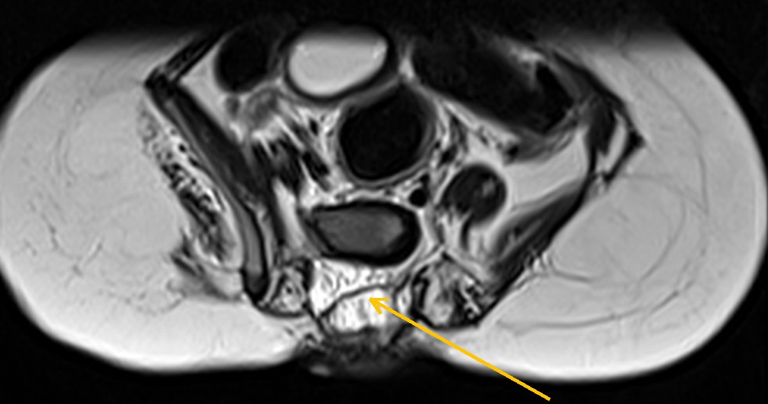
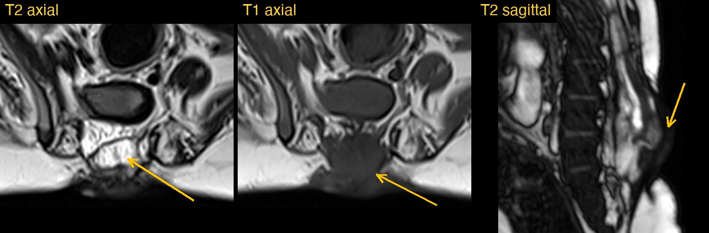
At the lumbosacral junction, where the lower back meets the sacrum, the normal layer of fat under the skin was replaced by a lobulated soft-tissue mass that pushed the skin outwards.
Why the brain belongs in a spine report
A defect at the base of the spine is easy to report on its own. But an open neural tube defect at this level almost always comes with changes in the brain: the back of the brain sits low and the fluid spaces enlarge. Reading only the requested region would leave half of the picture out.
How Monitraq's AI read the scan
- It described the defect: a lobulated, mixed-signal soft-tissue mass in the posterior midline at the lumbosacral junction, protruding beyond the bones at the back of the spine and replacing the fat under the skin. The bony arches at that level looked abnormally formed, and the skin contour bulged.
- It named the most likely cause: a lumbosacral posterior spinal dysraphism, favoured to be a myelomeningocele, with lipomyelomeningocele as the main alternative. It noted that this could be an open neural tube defect, and that the skin covering could not be confirmed on the images provided.
- It read the brain images too: marked enlargement of both lateral ventricles and the third ventricle, with a thinned brain mantle, which it reported as severe hydrocephalus. It noted that the lower cerebellum appeared to lie at or near the foramen magnum, with crowding at the junction of the skull and spine, and put these findings in keeping with the Chiari II spectrum.
- It flagged the scan for same-day radiologist review and listed the next steps: review the full lumbosacral sagittal and axial series to define the level of the defect, what the sac contains and whether skin covers it; correlate with any previous closure and shunt; perform a formal brain read with a midline sagittal view of the posterior fossa to assess the Chiari II features; and arrange neurosurgical correlation for the hydrocephalus.
Dr Priti, the radiologist who reviewed this case, reported an open neural tube defect: a lumbosacral myelomeningocele with Chiari II malformation and hydrocephalus.
What are a myelomeningocele and Chiari II malformation?
A myelomeningocele is the most common form of open spina bifida. During early pregnancy the neural tube, which becomes the spinal cord, fails to close at the bottom. The spinal cord and its coverings push out through a gap in the bones of the back. It is usually closed surgically, either soon after birth or before birth.
Almost every child with a myelomeningocele also has a Chiari II malformation: the cerebellum and brainstem sit lower than normal and crowd the opening at the base of the skull. This can block the flow of the fluid around the brain and cause hydrocephalus, which often needs a shunt. Taking folic acid before and in early pregnancy lowers the risk of neural tube defects.
A draft for the radiologist
A good radiologist reads the whole study, not only the region named on the request: a defect in the lower back means look at the brain. Monitraq's AI is built to do the same, and to show its reasoning so the radiologist can check it quickly.
It gives a draft, not a diagnosis. The AI flags, describes and suggests. A radiologist confirms, corrects and signs.
Monitraq's AI produces draft reports to support qualified radiologists. It is not a diagnosis and is not intended for use by patients. All case details have been removed or generalised to protect patient privacy, and the image has been cropped and stripped of identifying information.