Pleomorphic xanthoastrocytoma on MRI: how Monitraq's AI read it
This brain MRI shows a pleomorphic xanthoastrocytoma, a rare cortical-based tumour, as a large cystic and solid mass at the back of the right hemisphere. Here is what the scan showed, how Monitraq's AI described it, and why a radiologist signs every report.
MRI findings
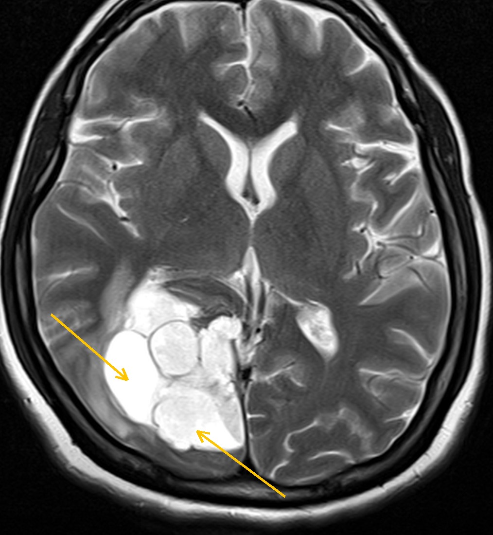
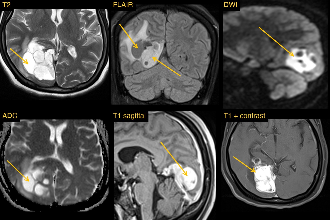
A patient had a full brain MRI with T2, FLAIR, diffusion, susceptibility, T1 and post-contrast T1 images. At the back of the right hemisphere, along its medial surface, in the medial parietal and occipital lobes, sat a large lobulated mass. It reached the falx in the midline and the tentorium below.
The mass was made of many compartments. Some were filled with very bright fluid on T2, divided by thin dark walls. Others were solid and much brighter on T1. Bright swelling (oedema) spread into the white matter around it, and part of the right lateral ventricle next to it was enlarged.
How Monitraq's AI read the scan
- It placed the mass: a lobulated, multiloculated mass in the right medial parieto-occipital lobe at the level of the right atrium (trigone) of the lateral ventricle, reaching the falx and tentorium. It measured about 45 x 50 mm on axial images.
- It described every part: very bright T2 locules with thin dark septa, dependent dark material suggesting fluid-blood levels, mixed FLAIR-bright solid parts and FLAIR-dark cystic parts, marked brightness on the sagittal T1 and the post-contrast T1, bright signal on DWI with mixed intermediate-to-high ADC, and a curvilinear dark rim on SWI.
- It was careful about what bright T1 means: without a matching T1 before contrast, it could not separate true enhancement from blood products, so it advised comparing the two or using subtraction images.
- It measured the effect on the brain: finger-like oedema into the right centrum semiovale, a midline shift under about 3 mm and no herniation. The right temporal horn and atrium were trapped and dilated (temporal horn about 10 mm wide), with possible fluid seeping into the tissue around it.
- It checked the tracts: on the colour DTI map, the right optic radiation looked displaced and oedematous, with the splenium preserved. It gave this low confidence.
- It gave a differential and flagged the scan: its favoured diagnosis was a haemorrhagic neoplasm (metastasis, high-grade glioma, or an intraventricular tumour such as atypical meningioma or ependymoma), with giant cavernoma and haemorrhage from a vascular malformation as the main alternatives. It asked for same-day radiologist review with neurosurgical input for the trapped temporal horn, and suggested SWI phase, spectroscopy review and angiography.
Dr Priti, the radiologist who reviewed this case, reported a pleomorphic xanthoastrocytoma: a cortical-based cystic and solid, avidly enhancing tumour of the right medial parieto-occipital lobe abutting the falx and tentorium.
The AI described the mass, its compartments, the oedema and the trapped ventricle in detail, and set out its own differential. Putting a name to a rare tumour from its MRI appearance takes expert judgement and clinical context, which is why a radiologist reviews and signs every report.
What is pleomorphic xanthoastrocytoma?
Pleomorphic xanthoastrocytoma (PXA) is a rare astrocytic brain tumour. It is usually found in children and young adults, and seizures are a common way for it to come to light. Its name describes cells that vary widely in shape (pleomorphic) and some that contain fat (xantho-).
PXA grows at the surface of the brain. It is based in the cortex and often touches the overlying meninges. The temporal lobe is the most common site, but any lobe can be involved. Most are lower grade, but some show anaplastic features and behave more aggressively.
Key MRI signs of pleomorphic xanthoastrocytoma
- A superficial, cortical-based mass, most often in the temporal lobe.
- A cyst with an enhancing solid nodule, with the nodule usually against the meninges.
- Strong enhancement of the solid part after contrast.
- Enhancement of the nearby meninges (a dural tail) in some cases.
- Oedema that is often mild compared with the size of the mass, though it varies.
Differential diagnosis
A cystic and solid cortical tumour has a short textbook list. Ganglioglioma is the closest mimic and also favours the temporal lobe in young patients. Pilocytic astrocytoma can show a cyst and nodule but sits more often in the cerebellum. Glioblastoma and metastasis are more likely in older adults, usually with more oedema and a thick, irregular enhancing rim. Blood products inside a mass add haemorrhagic tumours and cavernoma to the list.
Frequently asked questions
Which MRI sequence shows a pleomorphic xanthoastrocytoma best?
Post-contrast T1 shows the solid part and any enhancement of the nearby meninges best. T2 and FLAIR show the cyst and the surrounding oedema.
Where in the brain does a pleomorphic xanthoastrocytoma usually occur?
It is usually superficial and cortical-based, often reaching the meninges. The temporal lobe is the most common site, but the parietal, frontal and occipital lobes can also be involved.
Can MRI confirm a pleomorphic xanthoastrocytoma?
MRI can suggest it from the location and the cyst-and-nodule pattern. The diagnosis is confirmed by examining tissue under the microscope, often with testing for the BRAF V600E mutation.
A draft for the radiologist
A complex mass needs a careful, complete description before anyone names it. Monitraq's AI is built to give that description, flag the urgent parts and show its reasoning so the radiologist can check it quickly.
It gives a draft, not a diagnosis. The AI flags, describes and suggests. A radiologist confirms, corrects and signs.
Monitraq's AI produces draft reports to support qualified radiologists. It is not a diagnosis and is not intended for use by patients. All case details have been removed or generalised to protect patient privacy, and the images have been cropped and stripped of identifying information.